Minireviews Open Access
Copyright ©The Author(s) 2024. Published by Baishideng Publishing Group Inc. All rights reserved.
World J Crit Care Med. Jun 9, 2024; 13(2): 91212
Published online Jun 9, 2024. doi: 10.5492/wjccm.v13.i2.91212
Sound waves and solutions: Point-of-care ultrasonography for acute kidney injury in cirrhosis
David Aguirre-Villarreal, Departamento de Gastroenterología, Instituto Nacional de Ciencias Médicas y Nutrición Salvador Zubirán, Mexico City 14080, Mexico
Mario Andrés de Jesús Leal-Villarreal, Cardiología, Instituto Nacional de Cardiología Ignacio Chávez, Mexico City 14080, Mexico
Ignacio García-Juárez, Unidad de Hepatología y Trasplante, Departamento de Gastroenterología, Instituto Nacional de Ciencias Medicas y Nutricion Salvador Zubiran, Mexico City 14080, Mexico
Eduardo R Argaiz, Departamento de Nefrología y Metabolismo Mineral, Instituto Nacional de Ciencias Médicas y Nutrición Salvador Zubirán, Mexico City 14080, Mexico
Eduardo R Argaiz, Tecnológico de Monterrey, Escuela de Medicina y Ciencias de la Salud, Mexico City 64710, Mexico
Abhilash Koratala, Department of Nephrology, Medical College of Wisconsin, Milwaukee, WI 53226, United States
ORCID number: David Aguirre-Villarreal (0000-0001-8277-6846); Mario Andrés de Jesús Leal-Villarreal (0000-0003-2706-9545); Ignacio García-Juárez (0000-0003-2400-1887); Eduardo R Argaiz (0000-0002-5542-6098); Abhilash Koratala (0000-0001-5801-3574).
Author contributions: Aguirre-Villarreal D, Leal-Villarreal MAJ and Garcia-Juarez I drafted the initial version of the manuscript; Argaiz ER and Koratala A reviewed and revised the manuscript for critical intellectual content.
Supported by Research funding from KidneyCure and the American Society of Nephrology’s William and Sandra Bennett Clinical Scholars Grant (to Abhilash Koratala).
Conflict-of-interest statement: All authors report no conflicts of interest to declare.
Open-Access: This article is an open-access article that was selected by an in-house editor and fully peer-reviewed by external reviewers. It is distributed in accordance with the Creative Commons Attribution NonCommercial (CC BY-NC 4.0) license, which permits others to distribute, remix, adapt, build upon this work non-commercially, and license their derivative works on different terms, provided the original work is properly cited and the use is non-commercial. See: https://creativecommons.org/Licenses/by-nc/4.0/
Corresponding author: Abhilash Koratala, MD, Associate Professor, Department of Nephrology, Medical College of Wisconsin, 8701 W Watertown Plank Road, Room A 7633, Milwaukee, WI 53226, United States. akoratala@mcw.edu
Received: December 24, 2023
Revised: March 5, 2024
Accepted: April 22, 2024
Published online: June 9, 2024

Abstract

This article delves into the intricate challenges of acute kidney injury (AKI) in cirrhosis, a condition fraught with high morbidity and mortality. The complexities arise from distinguishing between various causes of AKI, particularly hemodynamic AKI, in cirrhotic patients, who experience hemodynamic changes due to portal hypertension. The term "hepatocardiorenal syndrome" is introduced to encapsulate the intricate interplay among the liver, heart, and kidneys. The narrative emphasizes the often-overlooked aspect of cardiac function in AKI assessments in cirrhosis, unveiling the prevalence of cirrhotic cardiomyopathy marked by impaired diastolic function. The conventional empiric approach involving volume expansion and vasopressors for hepatorenal syndrome is critically analyzed, highlighting potential risks and variable patient responses. We advocate for a nuanced algorithm for AKI evaluation in cirrhosis, prominently featuring point-of-care ultrasonography (POCUS). POCUS applications encompass assessing fluid tolerance, detecting venous congestion, and evaluating cardiac function.

Key Words: Point-of-care ultrasonography, Bedside ultrasound, Cirrhosis, Congestion, Acute kidney injury, Congestive nephropathy

Core Tip: In evaluating acute kidney injury in cirrhotic patients, point-of-care ultrasonography (POCUS) plays a pivotal role. This non-invasive diagnostic tool, when thoughtfully integrated, enhances sensitivity of physical examination, facilitates nuanced fluid management, and fine-tunes bedside hemodynamic evaluation. In the delicate interplay of liver, heart, and kidneys, POCUS emerges as an indispensable ally, refining assessments and steering towards optimal patient outcomes.


INTRODUCTION

Patients with cirrhosis frequently experience acute kidney injury (AKI) during hospitalization, and it is associated with a high morbidity and mortality[1,2]. Broadly, AKI can be secondary to renal hypoperfusion (hemodynamic AKI), intrinsic parenchymal damage or urinary obstruction and a systematic evaluation must be performed in all patients in order to ascertain its cause[3]. In patients with cirrhosis, it is particularly challenging to approach hemodynamic AKI, which can be caused by hypovolemia, distributive physiology (hepato-renal syndrome), intra-abdominal hypertension (IAH) or congestive nephropathy (type 1 cardio-renal syndrome).

HEMODYNAMIC CHANGES IN CIRRHOSIS

Hemodynamic changes in patients with cirrhosis and portal hypertension make them highly susceptible to AKI from renal hypoperfusion. Parenchymal distortion due regenerative nodules and fibrosis, as well as an increase in intrahepatic vascular tone are followed by resistance in portal inflow and portal hypertension[4]. In response, activation of vasodilators (mainly nitric oxide), results in systemic vasodilation and decreased systemic vascular resistance (SVR)[5,6]. Further, arteriovenous and portosystemic shunts can also contribute to a decrease in SVR[6]. Resulting hypoperfusion is initially compensated through the renin-angiotensin-aldosterone system (RAAS) and antidiuretic hormone mediated volume expansion, as well adrenergic activation[7,8]. These mechanisms increase cardiac output through increased preload and chronotropism[7,8]. However, if the resulting cardiac output proves to be insufficient for metabolic demand, and volume expansion exceeds the limits of the Frank-Starling mechanism, high-output cardiac failure (HOHF) will occur resulting in elevated filing pressures and venous congestion. HOHF is defined as congestive heart failure with elevated filling pressures in the setting of an elevated cardiac index (≥ 4 L/min/m2)[9]. Interestingly, cirrhosis was the second most prevalent cause of HOHF, surpassed only by obesity in a large case series[9]. Moreover, it has been recognized that compromised contractile reserve is common, even in individuals with maintained left ventricular ejection fraction (LVEF). Given that longitudinal contractile function tends to be impaired before radial function loss, recent guidelines propose utilizing global longitudinal strain, with a normal value of 18% or higher) to detect myocardial contractile dysfunction in those with preserved LVEF[10]. Therefore, although renal dysfunction can be aggravated by the splanchnic vasodilation and renal vasoconstriction, as in the case of the classical hepatorenal syndrome (HRS), it is also paramount to consider that decreased renal function could also be a consequence of venous congestion in the setting of advanced diastolic disfunction or even reduced cardiac output in the setting of reduced systolic function. The term hepatocardiorenal syndrome has been proposed to encompass the intricate interplay among the liver, heart, and kidneys[11].

HRS AND GUIDELINE-BASED TREATMENT

HRS is a potentially reversible reduction in glomerular filtration rate (GFR) that occurs in patients with advanced liver disease in the absence of shock, nephrotoxic drugs, and histological changes in renal parenchyma that does not respond to diuretic withdrawal and volume expansion[12]. It is thought that HRS is the consequence of the compensatory activation of the RAAS and sympathetic nervous system, with resulting renal vasoconstriction, suppression of vasodilatory mechanisms and thus a decreased GFR[13]. This is supported by reduced renal cortical perfusion on renal arteriography[14], the presence of high levels of renin activity, as well as high levels of norepinephrine in serum of patients with HRS[15]. Furthermore, the effectivity of terlipressin in some patients with HRS supports the role of splanchnic vasodilation as a cornerstone in its pathophysiology[16]. It is worth mentioning that other pathogenic factors contributing to HRS are systemic inflammation due to bacterial translocation, with release of pathogen-associated and damage-associated molecular patterns[17]. However, their role in HRS lies beyond the scope of this review. At present, HRS is a diagnosis of exclusion, and current guidelines recommend empiric treatment with volume expansion with albumin for 48 h to rule out volume depletion in patients with cirrhosis and AKI[18]. Persistent kidney injury after volume expansion is compatible with a diagnosis of HRS and vasopressors (terlipressin or norepinephrine) are the recommended treatment[13]. However, volume expansion and vasoconstrictor therapy are not without risks. Lessons learned from the ATTIRE trial are that routine albumin infusion in patients with decompensated cirrhosis is unbeneficial and associated with an increased risk of life-threatening adverse events, including fluid overload and respiratory failure due to pulmonary edema[19]. Moreover, higher rates of pulmonary failure were found in those who received both terlipressin and albumin in the CONFIRM trial[16], probably because of increased preload and afterload[20]. Hence, it is advisable to perform a thorough evaluation of volume status, possibly utilizing point-of-care ultrasonography (POCUS) before administering intravenous fluids[21-23].

CARDIAC FUNCTION IN ADVANCED CIRRHOSIS

The heterogeneous response to treatment might be a consequence of overlooking a key element of hemodynamics in patients with cirrhosis, which is cardiac function. Initially, a decreased SVR is compensated by volume expansion which is expected to result in increased cardiac output via the Frank-Starling mechanism. However, altered cardiac function, specifically impaired relaxation, can result in less tolerance for volume expansion resulting in increased filling pressures and venous congestion. Altered myocardial relaxation is frequently observed in patients with cirrhosis and this has been referred to as “cirrhotic cardiomyopathy”[10]. Cirrhotic cardiomyopathy is characterized by impaired diastolic function[24], enlarged cardiac chambers[25], a hampered response to stress[26], and electrophysiological changes in patients with advanced liver disease and no previous heart disease[27,28]. In a study by Ruíz-del-Árbol et al[29], 37 of 80 patients with cirrhosis had left ventricular diastolic dysfunction, which was associated with lower mean arterial pressures, higher Model for End-Stage Liver Disease scores, and greater risk of HRS-AKI and mortality. In 2019, the Cirrhotic Cardiomyopathy Consortium proposed echocardiographic criteria to define cirrhotic cardiomyopathy[10]. Under these criteria, the prevalence of cirrhotic cardiomyopathy has been reported to be as high as 60%[30].

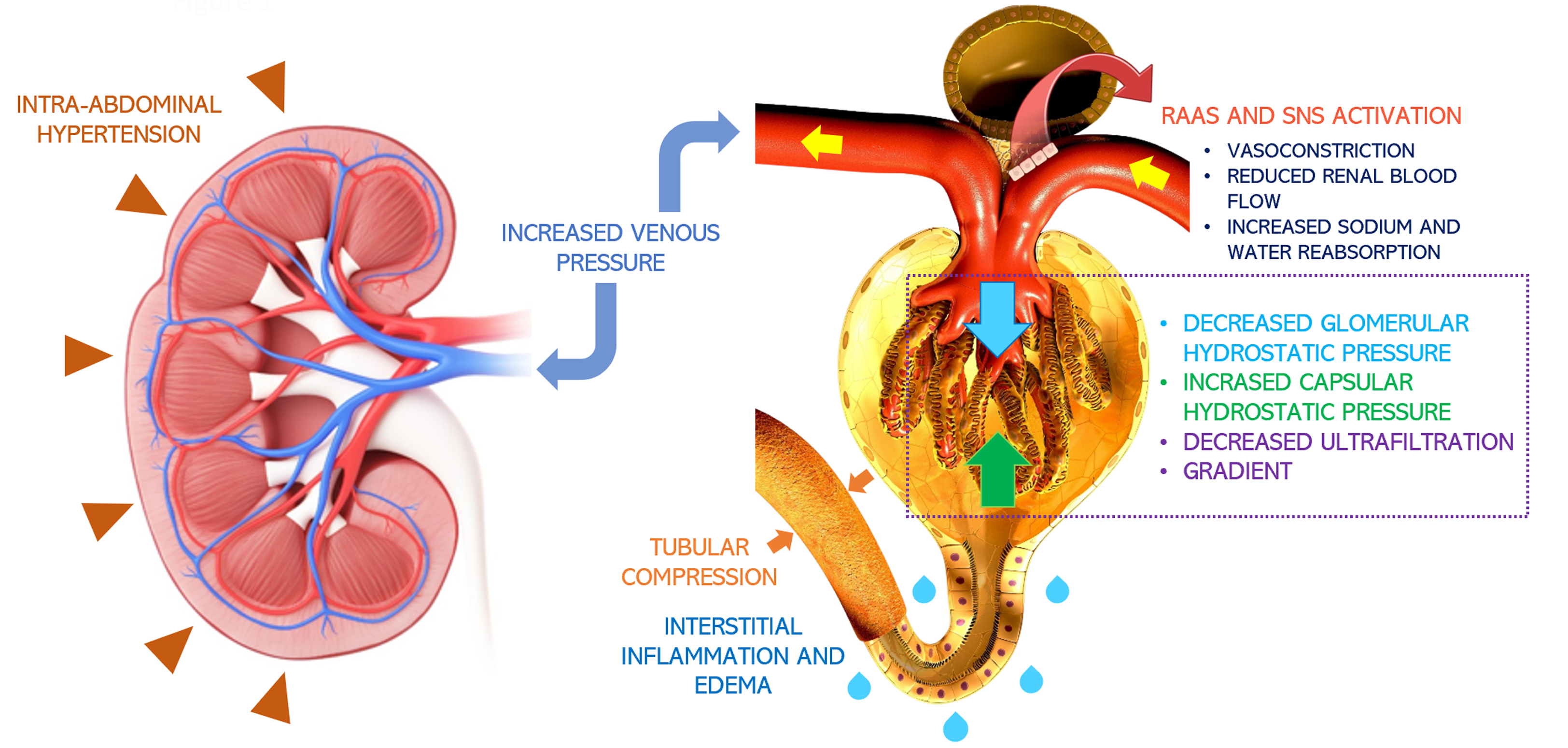
Failing to consider cardiac function in the setting of AKI in cirrhosis and treating with a “one-size-fits-all” approach could be detrimental. Increasing preload on an already surpassed Frank-Starling mechanism leads to further congestion, with no benefit on cardiac output or renal perfusion. Furthermore, the use of vasoconstrictors on patients with low contractility would increase the afterload on an already strained heart, which would further compromise cardiac output. In fact, we now know that venous congestion is the main hemodynamic determinant of renal dysfunction in patients with heart failure (Figure 1)[31]. AKI in the setting of severe venous congestion is likely to improve by achieving decongestion through diuresis, ultrafiltration and, possibly inotropic drugs in the setting of reduced contractility. Thus, it is imperative to perform an accurate hemodynamic evaluation in patients with cirrhosis and AKI to tailor their management. This approach is supported by a retrospective study by Pelayo et al[32], where 127 patients with cirrhosis and a diagnosis of HRS underwent a right heart catheterization, where 62% of patients were found with elevated pulmonary wedge pressures consistent with cardiorenal syndrome physiology[32]. These patients were treated with diuretics instead of volume expansion achieving adequate response.

Figure 1
Figure 1 Pathophysiology of congestive nephropathy: Congestion-induced acute renal dysfunction is mediated by retrograde transmission of central venous pressure to the kidneys, leading to development of interstitial edema, inflammation, and activation of the renin-angiotensin-aldosterone system and sympathetic nervous system[66]. This further results in global cessation of glomerular filtration. Intra-abdominal hypertension adds to the problem by simulating a tamponade pathophysiology together with increased interstitial pressures. Citation: Argaiz ER, Romero-Gonzalez G, Rola P, Spiegel R, Haycock KH, Koratala A. Bedside Ultrasound in the Management of Cardiorenal Syndromes: An Updated Review. Cardiorenal Med 2023; 13: 372-384. Copyright© The Author(s) 2023. Published by S. Karger AG, Basel, authors’ prior work published under CC BY-NC License.
INITIAL APPROACH TO AKI IN PATIENTS WITH CIRRHOSIS USING POCUS

HRS-AKI diagnosis mandates the exclusion of other causes of AKI, including shock, nephrotoxic drugs, and intrinsic renal damage. However, other potentially correctible causes of AKI such as obstructive AKI and abdominal compartment syndrome (ACS) are sometimes overlooked. Obstructive or post-renal AKI arises from an obstruction downstream of the renal collecting system, commonly due to urinary retention, benign prostatic hyperplasia, nephrolithiasis, malignancy or even an obstructed Foley catheter. Upon ultrasonographic examination, hydronephrosis and dilation of the pelvicalyceal system in suggestive of obstruction, and promptly relieving obstruction is warranted[33]. Tension ascites should also be considered, as IAH (defined as ≥ 12 mmHg) and ACS (defined as ≥ 20 mmHg and signs of organ dysfunction) reduce renal perfusion pressure and lead to AKI[34]. A large volume ascites and a small, collapsed inferior vena cava (IVC) with minimal respiratory variation should raise the suspicion for abdominal hypertension[35,36]. After ruling out obstruction and ACS, as well as shock, nephrotoxic drugs and intrinsic kidney damage, a systematic approach to hemodynamic AKI must be performed.

CAN POCUS ASSESS HEMODYNAMICS BETTER THAN PHYSICAL EXAMINATION?

Increased awareness of the risk of the overzealous administration of intravenous fluids have shifted paradigms towards the evaluation of fluid tolerance instead of fluid responsiveness[37]. Although fluid administration in true hypovolemia is indubitably beneficial, physical exam findings consistent with hypovolemia often lack sensitivity[37]. It is also important to realize that no ultrasonographic parameter can confirm the diagnosis of hypovolemia. Low cardiac filing pressures and absence of venous congestion do not indicate hypovolemia but rather the absence of congestive heart failure. However, the presence of pulmonary and systemic venous congestion is indicative of heart failure and volume intolerance[37]. Clinical evaluation of congestion is often inaccurate. In a study by Torino et al[38], the comparison of lung ultrasound with standardized lung auscultation revealed a low sensitivity (9%) of lung crackles in detecting extravascular lung water in end-stage renal disease. Similarly, Breidthardt et al[39] showed that in patients presenting with acute heart failure to the emergency department, treating physicians were unable to correctly assess the degree of congestion through clinical jugular vein examination when compared to invasive central venous pressure monitoring. Multiple studies have illustrated how POCUS enhances the sensitivity of conventional physical examination across various clinical contexts[40,41].

EVALUATION OF CONGESTION USING POCUS

POCUS has emerged as a non-invasive, bedside diagnostic tool that, when integrated with clinical and laboratory data, offers a holistic insight into a patient's hemodynamic status. This can substantially improve decision-making regarding fluid administration or removal by obtaining qualitative and quantitative data on cardiac function, as well as pulmonary and vascular congestion[42].

Right-sided congestion

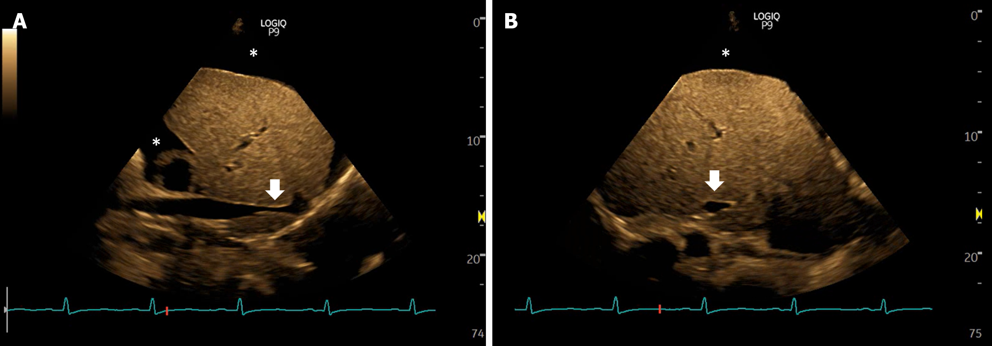
Right atrial pressure (RAP) serves as an estimation of preload, and an elevated RVP is suggestive of an absolute excess of venous return, or a relative one that surpasses cardiac output, which can then lead to congestive organ dysfunction through backward transmission. RAP can be invasively measured through catheterization or non-invasively estimated through POCUS. IVC ultrasound to assess size and respiratory variation has been widely adopted as a way to estimate RAP[43]. In a landmark study by Velez et al[44], authors performed IVC ultrasound in patients with cirrhosis and found that 21% had signs of elevated RAP (IVC diameter > 20 mm), these patients were treated with diuretics with adequate response. Despite this, it is important to recognize that IVC ultrasound in patients with cirrhosis faces important caveats. Hepatic fibrosis and the resulting stiffness can impair pressure-dependent changes in venous diameter[45]. Furthermore, IVC dilation can be secondary to factors unrelated to raised RAP, such as portosystemic collaterals draining to the IVC, constriction from an enlarged caudate lobe, or extension of hepatic vein thromboses or hepatocellular carcinoma into the IVC[46]. Importantly, IAH due to ascites can lead to IVC compression and nipping (Figure 2)[35]. A promising alternative to IVC POCUS to assess RAP is internal jugular vein (IJV) ultrasound. Previous studies in patients without cirrhosis have shown excellent correlation with RAP[43,47-49]. In patients with cirrhosis, IJV POCUS shows better correlation with RAP compared to IVC POCUS, possibly because changes in IJV diameter are independent of liver stiffness and IAH. Importantly, it can be performed even in patients with abundant ascites[50]. However, one should be cautious about caveats in IJV POCUS, including inappropriate head angle, excessive pressure on the vein with the transducer, inaccessibility due to previous thrombosis, variations in right atrial depth (as opposed to the traditional assumption of 5 cm), and the diversity of techniques in literature (e.g., respiratory variation in diameter, cross sectional area, response to Valsalva, column height, etc.).

Figure 2
Figure 2 Views of the inferior vena cava in a case of intra-abdominal hypertension showing narrowing or slit-like appearance of the intrahepatic portion (arrows), sometimes described as nipping. Asterisk indicates ascites. A: Long axis views; B: Transverse views.
POCUS in the assessment of renal congestion

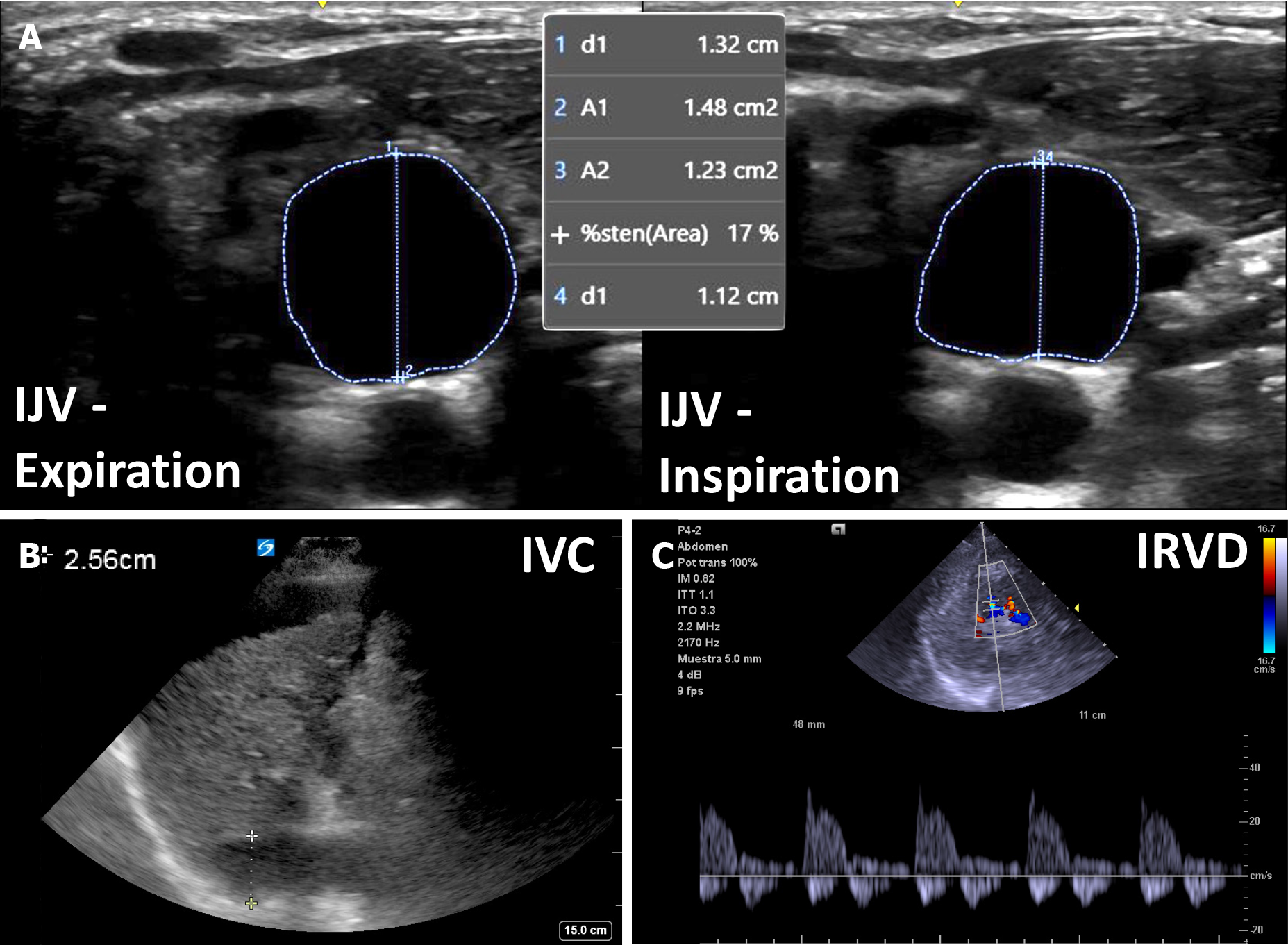
Unlike IVC/IJV POCUS, intrarenal venous doppler ultrasonography (IRVD) evaluates venous compliance of the renal vasculature. As RAP increases, increased volume and venous wall stretch lead to loss of venous compliance. This enhances distal transmission of pulsatile right atrial flow resulting in interrupted venous flow. Normal IRVD is continuous, while it becomes discontinuous with a biphasic or monophasic pattern in moderate and severe congestion, respectively[51]. Prospective studies have shown a strong independent association between congestive IRVD patterns and worse clinical outcomes in patients with heart failure[52] and pulmonary hypertension[53,54], likely mediated by congestion induced worsening renal function and diuretic resistance[55]. Altered IRVD predicts appropriate response diuretic treatment[56], and improvement in IRVD with diuretics is associated with improved clinical outcomes[57]. These data suggest that therapeutic efforts aimed at normalizing altered IRVD could be beneficial. However, intervention trials are needed. It is important to realize that no studies evaluating IRVD have been performed in patients with cirrhosis. However, it is our experience that it accurately reflects venous congestion in most cases. Hepatic and portal veins, commonly employed for assessing systemic venous congestion, may be unreliable in cirrhosis due to local structural and functional changes. Although not a universal observation[58], this area requires further study before making definitive recommendations. Figure 3 shows an example of venous congestion findings in cirrhosis.

Figure 3
Figure 3 An example of venous congestion in cirrhosis. A: Plethoric internal jugular vein with less than 25% antero-posterior inspiratory collapse; B: Plethoric inferior vena cava (> 2.0 cm); C: Biphasic intra-renal venous Doppler. IJV: Internal jugular vein; IVC: Inferior vena cava; IRVD: Intra-renal venous Doppler.
Left ventricular function, cardiac output, and left-sided congestion

In the setting of venous congestion, as suggested by a plethoric IVC/IJC, cardiac function should be evaluated. Albeit formal echocardiography warrants extensive training, focused cardiac ultrasonography (FoCUS) by non-echocardiographers might be enough to discern between normal, moderate, or severe systolic dysfunction. This was shown by Melamed et al[59], where intensivists with minimal training were able to estimate left ventricular (LV) function with reasonable accuracy when compared with experienced echocardiographers. Specifically, endocardial excursion, myocardial thickening, and septal motion of the anterior leaflet of the mitral valve are useful and feasible to obtain[60]. Moreover, stroke volume can be estimated by advanced POCUS users by measuring the velocity-time integral of the LV outflow tract[61]. Information obtained by FoCUS is crucial as a hyperdynamic circulation is better addressed using vasopressors while a severely depressed systolic function is unlikely to tolerate afterload increasing drugs such as terlipressin. Most patients with cirrhosis and heart failure will present with a preserved ejection fraction and diastolic dysfunction. While assessment of diastolic dysfunction is considered an advanced POCUS application, 6-point or 8-point lung ultrasound (LUS) reliably detects pulmonary congestion through the presence of B lines[62] and this is correlated with increased left ventricular end diastolic pressure[63]. The combination of albumin and terlipressin that is frequently initiated in patients with cirrhosis and AKI creates both an increase in preload and in afterload potentially driving hydrostatic pulmonary edema[64]. Acting upon LUS findings suggestive of pulmonary edema, which often precede clinical signs and symptoms could prevent further decline in respiratory function. In addition, LUS is effective in detecting pleural effusions. In a study of 116 patients with decompensated cirrhosis, anteroposterior chest X-rays missed about 40% of pleural effusions identified by LUS. Interestingly, detecting effusions with LUS was linked to a longer hospital stay (10 d vs 5.5 d, P < 0.001) and doubled mortality (39.7% vs 20.7%, P = 0.021)[65].

PROPOSED ALGORITHM

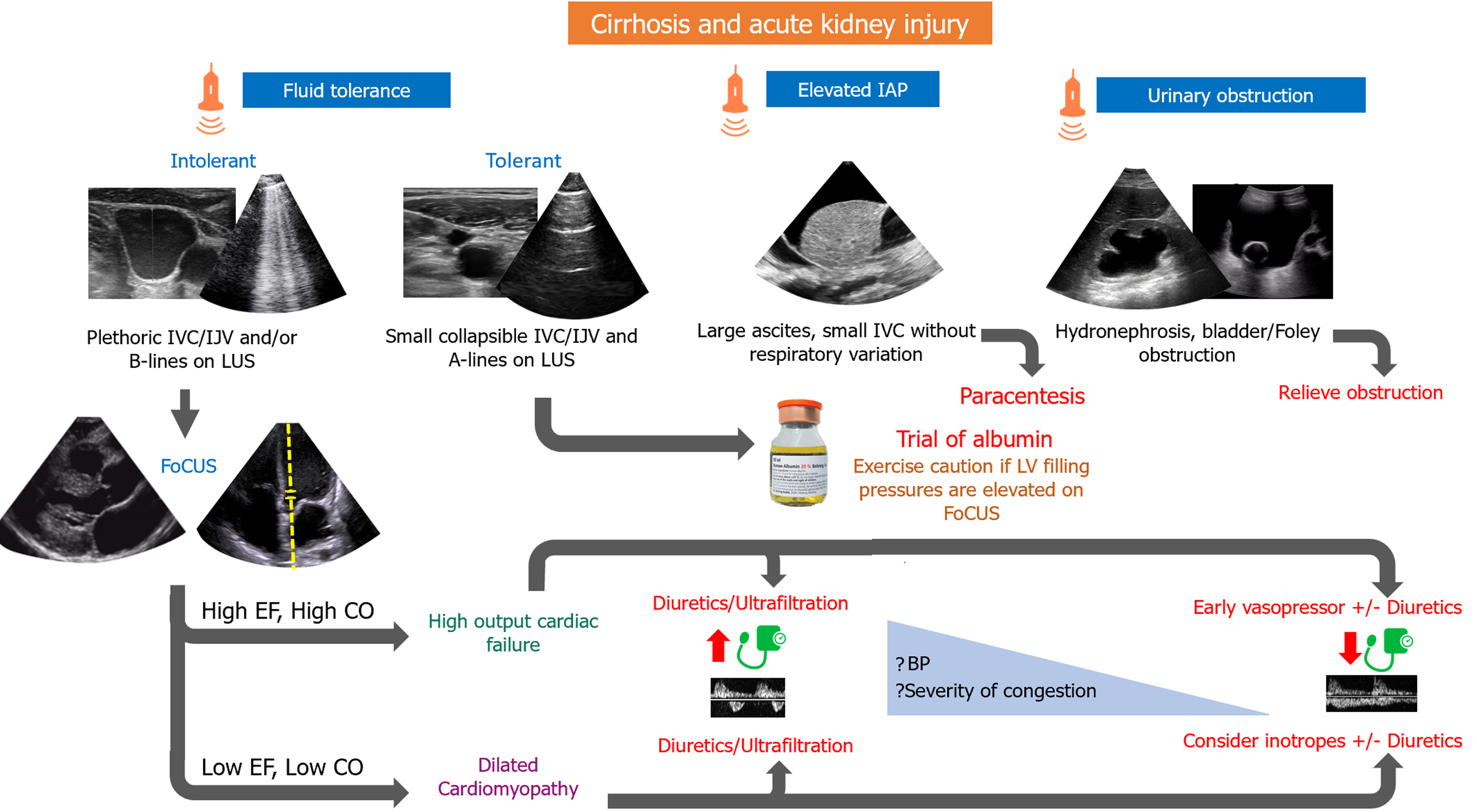
We propose that when evaluating a patient with AKI and cirrhosis, obstructive and intrinsic causes, as well as ACS, must initially be ruled out. In the case of hemodynamic AKI, fluid tolerance must be assessed through IJV/IVC POCUS, as well as LUS. If a patient is deemed fluid tolerant, an albumin trial could be a reasonable approach. In such cases, if FoCUS performed by an advanced user or a formal echocardiogram reveals Doppler stigmata of elevated left ventricular filling pressures, risks vs benefits of albumin therapy must be reweighed. On the other hand, if a patient is found to be fluid intolerant (pulmonary or venous congestion), then heart failure is diagnosed, and further cardiac evaluation should be performed. Most patients with cirrhosis and heart failure will display a hyperdynamic phenotype [high ejection fraction (EF), high cardiac output (CO)]. In these cases, decongestion should be attempted. Increased urine output and natriuresis can be achieved by means of diuretic drugs or by initiating vasopressor therapy in patients with inadequate mean arterial blood pressure. Vasopressors lead to increased renal blood flow and thus higher urine output. If congestion is severe, ultrafiltration might be needed in the presence of severe diuretic resistance (Figure 4).

Figure 4
Figure 4 Proposed algorithm for management of cirrhosis and acute kidney injury. IAP: Intra-abdominal pressure; IJV: Internal jugular vein; IVC: Inferior vena cava; LUS: Lung ultrasound; EF: Ejection fraction; CO: Cardiac output; LV: Left ventricular; BP: Blood pressure; FoCUS: Focused cardiac ultrasound.

This approach does not apply to patients who present with concomitant advanced cardiomyopathy and decreased contractility (low EF, low CO) where vasopressors could lead to further decrease in cardiac output. Besides diuretics/ultrafiltration, adequate decongestion may require inodilator drugs.

CONCLUSION

The blurred line between hepatorenal and cardiorenal syndrome is often difficult to discern, and hemodynamic AKI in patients with cirrhosis warrants a systematic evaluation of a patient´s individual physiology to provide a tailored approach to treatment as opposed to an empirical one. When evaluating AKI in cirrhosis, cardiac function is crucial but is frequently disregarded. A thorough hemodynamic assessment will aid in detecting those patients that do not benefit from routine HRS-AKI treatment with albumin and vasoconstrictors, and instead benefit from decongestion or inotropic drugs. Future investigations should consider incorporating multi-organ POCUS in the evaluation of these patients, moving away from isolated organ assessments. Prospective studies are needed to validate the proposed algorithm in larger cohorts and examine the impact of POCUS-guided interventions on patient outcomes. This approach should focus on understanding the effects of incorporating POCUS on tangible outcomes such as successful decongestion, clinical improvement, duration of intensive care unit stays, and hospitalization. It is important to note that expecting a mortality benefit solely from the use of a diagnostic test (POCUS) might be overly optimistic, given the limited therapies demonstrating mortality benefit in this patient population.

Having said that, it is crucial to exercise caution, as POCUS is operator-dependent, and effective patient management relies heavily on the operator’s skill in image acquisition, accurate interpretation, and clinical integration of data in the appropriate context. As such, it is necessary to have comprehensive training in POCUS across all levels of medical education, with robust quality assessment programs in place. There is an urgent need to establish global standards in POCUS training and competency assessment, and professional societies should collaborate to form multidisciplinary expert committees for this purpose.

Footnotes

Provenance and peer review: Invited article; Externally peer reviewed.

Peer-review model: Single blind

Specialty type: Critical care medicine

Country/Territory of origin: United States

Peer-review report’s classification

Scientific Quality: Grade B

Novelty: Grade B

Creativity or Innovation: Grade B

Scientific Significance: Grade B

P-Reviewer: Liu Y, China S-Editor: Che XX L-Editor: A P-Editor: Cai YX

References
1.  Belcher JM, Garcia-Tsao G, Sanyal AJ, Bhogal H, Lim JK, Ansari N, Coca SG, Parikh CR; TRIBE-AKI Consortium. Association of AKI with mortality and complications in hospitalized patients with cirrhosis. Hepatology. 2013;57:753-762.  [PubMed]  [DOI]  [Cited in This Article: ]  [Cited by in Crossref: 239]  [Cited by in F6Publishing: 249]  [Article Influence: 22.6]  [Reference Citation Analysis (0)]
2.  Desai AP, Knapp SM, Orman ES, Ghabril MS, Nephew LD, Anderson M, Ginès P, Chalasani NP, Patidar KR. Changing epidemiology and outcomes of acute kidney injury in hospitalized patients with cirrhosis - a US population-based study. J Hepatol. 2020;73:1092-1099.  [PubMed]  [DOI]  [Cited in This Article: ]  [Cited by in Crossref: 19]  [Cited by in F6Publishing: 40]  [Article Influence: 10.0]  [Reference Citation Analysis (0)]
3.  Velez JCQ. Hepatorenal Syndrome Type 1: From Diagnosis Ascertainment to Goal-Oriented Pharmacologic Therapy. Kidney360. 2022;3:382-395.  [PubMed]  [DOI]  [Cited in This Article: ]  [Cited by in Crossref: 6]  [Cited by in F6Publishing: 2]  [Article Influence: 1.0]  [Reference Citation Analysis (33)]
4.  Tapper EB, Parikh ND. Diagnosis and Management of Cirrhosis and Its Complications: A Review. JAMA. 2023;329:1589-1602.  [PubMed]  [DOI]  [Cited in This Article: ]  [Cited by in Crossref: 51]  [Cited by in F6Publishing: 39]  [Article Influence: 39.0]  [Reference Citation Analysis (33)]
5.  Martin PY, Ginès P, Schrier RW. Nitric oxide as a mediator of hemodynamic abnormalities and sodium and water retention in cirrhosis. N Engl J Med. 1998;339:533-541.  [PubMed]  [DOI]  [Cited in This Article: ]  [Cited by in Crossref: 310]  [Cited by in F6Publishing: 262]  [Article Influence: 10.1]  [Reference Citation Analysis (33)]
6.  Iwakiri Y, Groszmann RJ. The hyperdynamic circulation of chronic liver diseases: from the patient to the molecule. Hepatology. 2006;43:S121-S131.  [PubMed]  [DOI]  [Cited in This Article: ]  [Cited by in Crossref: 418]  [Cited by in F6Publishing: 374]  [Article Influence: 20.8]  [Reference Citation Analysis (14)]
7.  Bernardi M, Trevisani F, Gasbarrini A, Gasbarrini G. Hepatorenal disorders: role of the renin-angiotensin-aldosterone system. Semin Liver Dis. 1994;14:23-34.  [PubMed]  [DOI]  [Cited in This Article: ]  [Cited by in Crossref: 53]  [Cited by in F6Publishing: 52]  [Article Influence: 1.7]  [Reference Citation Analysis (34)]
8.  Henriksen JH, Møller S, Ring-Larsen H, Christensen NJ. The sympathetic nervous system in liver disease. J Hepatol. 1998;29:328-341.  [PubMed]  [DOI]  [Cited in This Article: ]  [Cited by in Crossref: 106]  [Cited by in F6Publishing: 112]  [Article Influence: 4.3]  [Reference Citation Analysis (33)]
9.  Reddy YNV, Melenovsky V, Redfield MM, Nishimura RA, Borlaug BA. High-Output Heart Failure: A 15-Year Experience. J Am Coll Cardiol. 2016;68:473-482.  [PubMed]  [DOI]  [Cited in This Article: ]  [Cited by in Crossref: 141]  [Cited by in F6Publishing: 113]  [Article Influence: 14.1]  [Reference Citation Analysis (1)]
10.  Izzy M, VanWagner LB, Lin G, Altieri M, Findlay JY, Oh JK, Watt KD, Lee SS; Cirrhotic Cardiomyopathy Consortium. Redefining Cirrhotic Cardiomyopathy for the Modern Era. Hepatology. 2020;71:334-345.  [PubMed]  [DOI]  [Cited in This Article: ]  [Cited by in Crossref: 119]  [Cited by in F6Publishing: 178]  [Article Influence: 44.5]  [Reference Citation Analysis (1)]
11.  Kazory A, Ronco C. Hepatorenal Syndrome or Hepatocardiorenal Syndrome: Revisiting Basic Concepts in View of Emerging Data. Cardiorenal Med. 2019;9:1-7.  [PubMed]  [DOI]  [Cited in This Article: ]  [Cited by in Crossref: 25]  [Cited by in F6Publishing: 25]  [Article Influence: 4.2]  [Reference Citation Analysis (1)]
12.  Angeli P, Gines P, Wong F, Bernardi M, Boyer TD, Gerbes A, Moreau R, Jalan R, Sarin SK, Piano S, Moore K, Lee SS, Durand F, Salerno F, Caraceni P, Kim WR, Arroyo V, Garcia-Tsao G; International Club of Ascites. Diagnosis and management of acute kidney injury in patients with cirrhosis: revised consensus recommendations of the International Club of Ascites. Gut. 2015;64:531-537.  [PubMed]  [DOI]  [Cited in This Article: ]  [Cited by in Crossref: 278]  [Cited by in F6Publishing: 320]  [Article Influence: 35.6]  [Reference Citation Analysis (1)]
13.  Garcia-Tsao G, Abraldes JG, Rich NE, Wong VW. AGA Clinical Practice Update on the Use of Vasoactive Drugs and Intravenous Albumin in Cirrhosis: Expert Review. Gastroenterology. 2024;166:202-210.  [PubMed]  [DOI]  [Cited in This Article: ]  [Cited by in Crossref: 8]  [Cited by in F6Publishing: 1]  [Article Influence: 1.0]  [Reference Citation Analysis (1)]
14.  Epstein M, Berk DP, Hollenberg NK, Adams DF, Chalmers TC, Abrams HL, Merrill JP. Renal failure in the patient with cirrhosis. The role of active vasoconstriction. Am J Med. 1970;49:175-185.  [PubMed]  [DOI]  [Cited in This Article: ]  [Cited by in Crossref: 328]  [Cited by in F6Publishing: 277]  [Article Influence: 5.1]  [Reference Citation Analysis (0)]
15.  Ruiz-del-Arbol L, Monescillo A, Arocena C, Valer P, Ginès P, Moreira V, Milicua JM, Jiménez W, Arroyo V. Circulatory function and hepatorenal syndrome in cirrhosis. Hepatology. 2005;42:439-447.  [PubMed]  [DOI]  [Cited in This Article: ]  [Cited by in Crossref: 412]  [Cited by in F6Publishing: 369]  [Article Influence: 19.4]  [Reference Citation Analysis (0)]
16.  Wong F, Pappas SC, Curry MP, Reddy KR, Rubin RA, Porayko MK, Gonzalez SA, Mumtaz K, Lim N, Simonetto DA, Sharma P, Sanyal AJ, Mayo MJ, Frederick RT, Escalante S, Jamil K; CONFIRM Study Investigators. Terlipressin plus Albumin for the Treatment of Type 1 Hepatorenal Syndrome. N Engl J Med. 2021;384:818-828.  [PubMed]  [DOI]  [Cited in This Article: ]  [Cited by in Crossref: 121]  [Cited by in F6Publishing: 217]  [Article Influence: 72.3]  [Reference Citation Analysis (0)]
17.  Ginès P, Solà E, Angeli P, Wong F, Nadim MK, Kamath PS. Hepatorenal syndrome. Nat Rev Dis Primers. 2018;4:23.  [PubMed]  [DOI]  [Cited in This Article: ]  [Cited by in Crossref: 110]  [Cited by in F6Publishing: 142]  [Article Influence: 23.7]  [Reference Citation Analysis (1)]
18.  Flamm SL, Wong F, Ahn J, Kamath PS. AGA Clinical Practice Update on the Evaluation and Management of Acute Kidney Injury in Patients With Cirrhosis: Expert Review. Clin Gastroenterol Hepatol. 2022;20:2707-2716.  [PubMed]  [DOI]  [Cited in This Article: ]  [Cited by in Crossref: 24]  [Cited by in F6Publishing: 24]  [Article Influence: 12.0]  [Reference Citation Analysis (0)]
19.  China L, Freemantle N, Forrest E, Kallis Y, Ryder SD, Wright G, Portal AJ, Becares Salles N, Gilroy DW, O'Brien A; ATTIRE Trial Investigators. A Randomized Trial of Albumin Infusions in Hospitalized Patients with Cirrhosis. N Engl J Med. 2021;384:808-817.  [PubMed]  [DOI]  [Cited in This Article: ]  [Cited by in Crossref: 112]  [Cited by in F6Publishing: 156]  [Article Influence: 52.0]  [Reference Citation Analysis (0)]
20.  Garcia-Tsao G. Terlipressin and Intravenous Albumin in Advanced Cirrhosis - Friend and Foe. N Engl J Med. 2021;384:869-871.  [PubMed]  [DOI]  [Cited in This Article: ]  [Cited by in Crossref: 4]  [Cited by in F6Publishing: 5]  [Article Influence: 1.7]  [Reference Citation Analysis (0)]
21.  Nadim MK, Garcia-Tsao G. Acute Kidney Injury in Patients with Cirrhosis. N Engl J Med. 2023;388:733-745.  [PubMed]  [DOI]  [Cited in This Article: ]  [Cited by in F6Publishing: 39]  [Reference Citation Analysis (0)]
22.  Koratala A, Reisinger N. Point of Care Ultrasound in Cirrhosis-Associated Acute Kidney Injury: Beyond Inferior Vena Cava. Kidney360. 2022;3:1965-1968.  [PubMed]  [DOI]  [Cited in This Article: ]  [Cited by in F6Publishing: 9]  [Reference Citation Analysis (0)]
23.  Koratala A, Ronco C, Kazory A. Albumin Infusion in Patients with Cirrhosis: Time for POCUS-Enhanced Physical Examination. Cardiorenal Med. 2021;11:161-165.  [PubMed]  [DOI]  [Cited in This Article: ]  [Cited by in Crossref: 4]  [Cited by in F6Publishing: 4]  [Article Influence: 1.3]  [Reference Citation Analysis (0)]
24.  Finucci G, Desideri A, Sacerdoti D, Bolognesi M, Merkel C, Angeli P, Gatta A. Left ventricular diastolic function in liver cirrhosis. Scand J Gastroenterol. 1996;31:279-284.  [PubMed]  [DOI]  [Cited in This Article: ]  [Cited by in Crossref: 100]  [Cited by in F6Publishing: 88]  [Article Influence: 3.1]  [Reference Citation Analysis (0)]
25.  Pozzi M, Carugo S, Boari G, Pecci V, de Ceglia S, Maggiolini S, Bolla GB, Roffi L, Failla M, Grassi G, Giannattasio C, Mancia G. Evidence of functional and structural cardiac abnormalities in cirrhotic patients with and without ascites. Hepatology. 1997;26:1131-1137.  [PubMed]  [DOI]  [Cited in This Article: ]  [Cited by in Crossref: 15]  [Cited by in F6Publishing: 57]  [Article Influence: 2.1]  [Reference Citation Analysis (0)]
26.  Wong F, Girgrah N, Graba J, Allidina Y, Liu P, Blendis L. The cardiac response to exercise in cirrhosis. Gut. 2001;49:268-275.  [PubMed]  [DOI]  [Cited in This Article: ]  [Cited by in Crossref: 189]  [Cited by in F6Publishing: 188]  [Article Influence: 8.2]  [Reference Citation Analysis (0)]
27.  Wong F. Cirrhotic cardiomyopathy. Hepatol Int. 2009;3:294-304.  [PubMed]  [DOI]  [Cited in This Article: ]  [Cited by in Crossref: 129]  [Cited by in F6Publishing: 128]  [Article Influence: 8.0]  [Reference Citation Analysis (0)]
28.  Møller S, Henriksen JH. Cardiovascular complications of cirrhosis. Gut. 2008;57:268-278.  [PubMed]  [DOI]  [Cited in This Article: ]  [Cited by in Crossref: 279]  [Cited by in F6Publishing: 299]  [Article Influence: 18.7]  [Reference Citation Analysis (0)]
29.  Ruíz-del-Árbol L, Achécar L, Serradilla R, Rodríguez-Gandía MÁ, Rivero M, Garrido E, Natcher JJ. Diastolic dysfunction is a predictor of poor outcomes in patients with cirrhosis, portal hypertension, and a normal creatinine. Hepatology. 2013;58:1732-1741.  [PubMed]  [DOI]  [Cited in This Article: ]  [Cited by in Crossref: 107]  [Cited by in F6Publishing: 108]  [Article Influence: 9.8]  [Reference Citation Analysis (0)]
30.  Razpotnik M, Bota S, Wimmer P, Hackl M, Lesnik G, Alber H, Peck-Radosavljevic M. The prevalence of cirrhotic cardiomyopathy according to different diagnostic criteria. Liver Int. 2021;41:1058-1069.  [PubMed]  [DOI]  [Cited in This Article: ]  [Cited by in Crossref: 14]  [Cited by in F6Publishing: 25]  [Article Influence: 8.3]  [Reference Citation Analysis (0)]
31.  Mullens W, Abrahams Z, Francis GS, Sokos G, Taylor DO, Starling RC, Young JB, Tang WHW. Importance of venous congestion for worsening of renal function in advanced decompensated heart failure. J Am Coll Cardiol. 2009;53:589-596.  [PubMed]  [DOI]  [Cited in This Article: ]  [Cited by in Crossref: 1048]  [Cited by in F6Publishing: 1075]  [Article Influence: 71.7]  [Reference Citation Analysis (0)]
32.  Pelayo J, Lo KB, Sultan S, Quintero E, Peterson E, Salacupa G, Zanoria MA, Guarin G, Helfman B, Sanon J, Mathew R, Yazdanyar A, Navarro V, Pressman G, Rangaswami J. Invasive hemodynamic parameters in patients with hepatorenal syndrome. Int J Cardiol Heart Vasc. 2022;42:101094.  [PubMed]  [DOI]  [Cited in This Article: ]  [Reference Citation Analysis (0)]
33.  Moses AA, Fernandez HE. Ultrasonography in Acute Kidney Injury. POCUS J. 2022;7:35-44.  [PubMed]  [DOI]  [Cited in This Article: ]  [Cited by in Crossref: 2]  [Cited by in F6Publishing: 3]  [Article Influence: 1.5]  [Reference Citation Analysis (0)]
34.  Chang Y, Qi X, Li Z, Wang F, Wang S, Zhang Z, Xiao C, Ding T, Yang C. Hepatorenal syndrome: insights into the mechanisms of intra-abdominal hypertension. Int J Clin Exp Pathol. 2013;6:2523-2528.  [PubMed]  [DOI]  [Cited in This Article: ]
35.  Wachsberg RH. Narrowing of the upper abdominal inferior vena cava in patients with elevated intraabdominal pressure: sonographic observations. J Ultrasound Med. 2000;19:217-222.  [PubMed]  [DOI]  [Cited in This Article: ]  [Cited by in Crossref: 19]  [Cited by in F6Publishing: 20]  [Article Influence: 0.8]  [Reference Citation Analysis (0)]
36.  de Jesús Leal-Villarreal MA, Argaiz E. Assessment of venous congestion in abdominal compartment syndrome. Eur Heart J Case Rep. 2023;7:ytad271.  [PubMed]  [DOI]  [Cited in This Article: ]  [Reference Citation Analysis (0)]
37.  Argaiz ER, Rola P, Haycock KH, Verbrugge FH. Fluid management in acute kidney injury: from evaluating fluid responsiveness towards assessment of fluid tolerance. Eur Heart J Acute Cardiovasc Care. 2022;11:786-793.  [PubMed]  [DOI]  [Cited in This Article: ]  [Cited by in Crossref: 3]  [Reference Citation Analysis (0)]
38.  Torino C, Gargani L, Sicari R, Letachowicz K, Ekart R, Fliser D, Covic A, Siamopoulos K, Stavroulopoulos A, Massy ZA, Fiaccadori E, Caiazza A, Bachelet T, Slotki I, Martinez-Castelao A, Coudert-Krier MJ, Rossignol P, Gueler F, Hannedouche T, Panichi V, Wiecek A, Pontoriero G, Sarafidis P, Klinger M, Hojs R, Seiler-Mussler S, Lizzi F, Siriopol D, Balafa O, Shavit L, Tripepi R, Mallamaci F, Tripepi G, Picano E, London GM, Zoccali C. The Agreement between Auscultation and Lung Ultrasound in Hemodialysis Patients: The LUST Study. Clin J Am Soc Nephrol. 2016;11:2005-2011.  [PubMed]  [DOI]  [Cited in This Article: ]  [Cited by in Crossref: 100]  [Cited by in F6Publishing: 115]  [Article Influence: 14.4]  [Reference Citation Analysis (0)]
39.  Breidthardt T, Moreno-Weidmann Z, Uthoff H, Sabti Z, Aeppli S, Puelacher C, Stallone F, Twerenbold R, Wildi K, Kozhuharov N, Wussler D, Flores D, Shrestha S, Badertscher P, Boeddinghaus J, Nestelberger T, Gimenez MR, Staub D, Aschwanden M, Lohrmann J, Pfister O, Osswald S, Mueller C. How accurate is clinical assessment of neck veins in the estimation of central venous pressure in acute heart failure? Insights from a prospective study. Eur J Heart Fail. 2018;20:1160-1162.  [PubMed]  [DOI]  [Cited in This Article: ]  [Cited by in Crossref: 10]  [Cited by in F6Publishing: 10]  [Article Influence: 1.7]  [Reference Citation Analysis (0)]
40.  Koratala A, Kazory A. An Introduction to Point-of-Care Ultrasound: Laennec to Lichtenstein. Adv Chronic Kidney Dis. 2021;28:193-199.  [PubMed]  [DOI]  [Cited in This Article: ]  [Cited by in Crossref: 3]  [Cited by in F6Publishing: 10]  [Article Influence: 3.3]  [Reference Citation Analysis (0)]
41.  Bhagra A, Tierney DM, Sekiguchi H, Soni NJ. Point-of-Care Ultrasonography for Primary Care Physicians and General Internists. Mayo Clin Proc. 2016;91:1811-1827.  [PubMed]  [DOI]  [Cited in This Article: ]  [Cited by in Crossref: 115]  [Cited by in F6Publishing: 134]  [Article Influence: 16.8]  [Reference Citation Analysis (0)]
42.  Argaiz ER, Koratala A, Reisinger N. Comprehensive Assessment of Fluid Status by Point-of-Care Ultrasonography. Kidney360. 2021;2:1326-1338.  [PubMed]  [DOI]  [Cited in This Article: ]  [Cited by in Crossref: 29]  [Cited by in F6Publishing: 23]  [Article Influence: 7.7]  [Reference Citation Analysis (0)]
43.  Kircher BJ, Himelman RB, Schiller NB. Noninvasive estimation of right atrial pressure from the inspiratory collapse of the inferior vena cava. Am J Cardiol. 1990;66:493-496.  [PubMed]  [DOI]  [Cited in This Article: ]  [Cited by in Crossref: 754]  [Cited by in F6Publishing: 717]  [Article Influence: 21.1]  [Reference Citation Analysis (0)]
44.  Velez JCQ, Petkovich B, Karakala N, Huggins JT. Point-of-Care Echocardiography Unveils Misclassification of Acute Kidney Injury as Hepatorenal Syndrome. Am J Nephrol. 2019;50:204-211.  [PubMed]  [DOI]  [Cited in This Article: ]  [Cited by in Crossref: 21]  [Cited by in F6Publishing: 47]  [Article Influence: 9.4]  [Reference Citation Analysis (0)]
45.  Kitamura H, Kobayashi C. Impairment of change in diameter of the hepatic portion of the inferior vena cava: a sonographic sign of liver fibrosis or cirrhosis. J Ultrasound Med. 2005;24:355-359; quiz 360.  [PubMed]  [DOI]  [Cited in This Article: ]  [Cited by in Crossref: 20]  [Cited by in F6Publishing: 9]  [Article Influence: 0.5]  [Reference Citation Analysis (0)]
46.  Wachsberg RH, Levine CD, Maldjian PD, Simmons MZ. Dilatation of the inferior vena cava in patients with cirrhotic portal hypertension. Causes and imaging findings. Clin Imaging. 1998;22:48-53.  [PubMed]  [DOI]  [Cited in This Article: ]  [Cited by in Crossref: 7]  [Cited by in F6Publishing: 6]  [Article Influence: 0.2]  [Reference Citation Analysis (0)]
47.  Arthur ME, Landolfo C, Wade M, Castresana MR. Inferior vena cava diameter (IVCD) measured with transesophageal echocardiography (TEE) can be used to derive the central venous pressure (CVP) in anesthetized mechanically ventilated patients. Echocardiography. 2009;26:140-149.  [PubMed]  [DOI]  [Cited in This Article: ]  [Cited by in Crossref: 36]  [Cited by in F6Publishing: 42]  [Article Influence: 2.8]  [Reference Citation Analysis (0)]
48.  Mesin L, Albani S, Sinagra G. Non-invasive Estimation of Right Atrial Pressure Using Inferior Vena Cava Echography. Ultrasound Med Biol. 2019;45:1331-1337.  [PubMed]  [DOI]  [Cited in This Article: ]  [Cited by in Crossref: 12]  [Cited by in F6Publishing: 9]  [Article Influence: 1.8]  [Reference Citation Analysis (0)]
49.  Longino A, Martin K, Leyba K, Siegel G, Gill E, Douglas IS, Burke J. Correlation between the VExUS score and right atrial pressure: a pilot prospective observational study. Crit Care. 2023;27:205.  [PubMed]  [DOI]  [Cited in This Article: ]  [Cited by in Crossref: 23]  [Cited by in F6Publishing: 17]  [Article Influence: 17.0]  [Reference Citation Analysis (0)]
50.  Leal-Villarreal MAJ, Aguirre-Villarreal D, Vidal-Mayo JJ, Argaiz ER, García-Juárez I. Correlation of Internal Jugular Vein Collapsibility With Central Venous Pressure in Patients With Liver Cirrhosis. Am J Gastroenterol. 2023;118:1684-1687.  [PubMed]  [DOI]  [Cited in This Article: ]  [Cited by in Crossref: 3]  [Cited by in F6Publishing: 3]  [Article Influence: 3.0]  [Reference Citation Analysis (0)]
51.  Argaiz ER. VExUS Nexus: Bedside Assessment of Venous Congestion. Adv Chronic Kidney Dis. 2021;28:252-261.  [PubMed]  [DOI]  [Cited in This Article: ]  [Cited by in Crossref: 2]  [Cited by in F6Publishing: 22]  [Article Influence: 7.3]  [Reference Citation Analysis (0)]
52.  Iida N, Seo Y, Sai S, Machino-Ohtsuka T, Yamamoto M, Ishizu T, Kawakami Y, Aonuma K. Clinical Implications of Intrarenal Hemodynamic Evaluation by Doppler Ultrasonography in Heart Failure. JACC Heart Fail. 2016;4:674-682.  [PubMed]  [DOI]  [Cited in This Article: ]  [Cited by in Crossref: 121]  [Cited by in F6Publishing: 183]  [Article Influence: 22.9]  [Reference Citation Analysis (0)]
53.  Husain-Syed F, Birk HW, Ronco C, Schörmann T, Tello K, Richter MJ, Wilhelm J, Sommer N, Steyerberg E, Bauer P, Walmrath HD, Seeger W, McCullough PA, Gall H, Ghofrani HA. Doppler-Derived Renal Venous Stasis Index in the Prognosis of Right Heart Failure. J Am Heart Assoc. 2019;8:e013584.  [PubMed]  [DOI]  [Cited in This Article: ]  [Cited by in Crossref: 28]  [Cited by in F6Publishing: 54]  [Article Influence: 10.8]  [Reference Citation Analysis (0)]
54.  Gómez-Rodríguez C, Tadeo-Espinoza H, Solis-Huerta F, Leal-Villarreal MAJ, Guerrero-Cabrera P, Cruz N, Gaytan-Arocha JE, Soto-Mota A, Vasquez Z, Gamba G, Verbrugge FH, Argaiz ER. Hemodynamic Evaluation of Right-Sided Congestion With Doppler Ultrasonography in Pulmonary Hypertension. Am J Cardiol. 2023;203:459-462.  [PubMed]  [DOI]  [Cited in This Article: ]  [Reference Citation Analysis (0)]
55.  Nijst P, Martens P, Dupont M, Tang WHW, Mullens W. Intrarenal Flow Alterations During Transition From Euvolemia to Intravascular Volume Expansion in Heart Failure Patients. JACC Heart Fail. 2017;5:672-681.  [PubMed]  [DOI]  [Cited in This Article: ]  [Cited by in Crossref: 66]  [Cited by in F6Publishing: 69]  [Article Influence: 9.9]  [Reference Citation Analysis (0)]
56.  Guinot PG, Bahr PA, Andrei S, Popescu BA, Caruso V, Mertes PM, Berthoud V, Nguyen M, Bouhemad B. Doppler study of portal vein and renal venous velocity predict the appropriate fluid response to diuretic in ICU: a prospective observational echocardiographic evaluation. Crit Care. 2022;26:305.  [PubMed]  [DOI]  [Cited in This Article: ]  [Cited by in F6Publishing: 9]  [Reference Citation Analysis (0)]
57.  Turrini F, Galassi M, Sacchi A, Ricco' B, Chester J, Famiglietti E, Messora R, Bertolotti M, Pinelli G. Intrarenal Venous Doppler as a novel marker for optimal decongestion, patient management, and prognosis in Acute Decompensated Heart Failure. Eur Heart J Acute Cardiovasc Care. 2023;12:673-681.  [PubMed]  [DOI]  [Cited in This Article: ]  [Reference Citation Analysis (0)]
58.  Koratala A, Taleb Abdellah A, Reisinger N. Nephrologist-performed point-of-care venous excess Doppler ultrasound (VExUS) in the management of acute kidney injury. J Ultrasound. 2023;26:301-306.  [PubMed]  [DOI]  [Cited in This Article: ]  [Reference Citation Analysis (0)]
59.  Melamed R, Sprenkle MD, Ulstad VK, Herzog CA, Leatherman JW. Assessment of left ventricular function by intensivists using hand-held echocardiography. Chest. 2009;135:1416-1420.  [PubMed]  [DOI]  [Cited in This Article: ]  [Cited by in Crossref: 159]  [Cited by in F6Publishing: 166]  [Article Influence: 11.1]  [Reference Citation Analysis (0)]
60.  Andrus P, Dean A. Focused cardiac ultrasound. Glob Heart. 2013;8:299-303.  [PubMed]  [DOI]  [Cited in This Article: ]  [Cited by in Crossref: 11]  [Cited by in F6Publishing: 13]  [Article Influence: 1.2]  [Reference Citation Analysis (0)]
61.  Blanco P. Rationale for using the velocity-time integral and the minute distance for assessing the stroke volume and cardiac output in point-of-care settings. Ultrasound J. 2020;12:21.  [PubMed]  [DOI]  [Cited in This Article: ]  [Cited by in Crossref: 30]  [Cited by in F6Publishing: 67]  [Article Influence: 16.8]  [Reference Citation Analysis (0)]
62.  Li N, Zhu Y, Zeng J. Clinical value of pulmonary congestion detection by lung ultrasound in patients with chronic heart failure. Clin Cardiol. 2021;44:1488-1496.  [PubMed]  [DOI]  [Cited in This Article: ]  [Cited by in Crossref: 5]  [Cited by in F6Publishing: 7]  [Article Influence: 2.3]  [Reference Citation Analysis (0)]
63.  Imanishi J, Maeda T, Ujiro S, Masuda M, Kusakabe Y, Takemoto M, Fujimoto W, Kuroda K, Yamashita S, Iwasaki M, Todoroki T, Okuda M. Association between B-lines on lung ultrasound, invasive haemodynamics, and prognosis in acute heart failure patients. Eur Heart J Acute Cardiovasc Care. 2023;12:115-123.  [PubMed]  [DOI]  [Cited in This Article: ]  [Cited by in F6Publishing: 6]  [Reference Citation Analysis (0)]
64.  Allegretti AS, Subramanian RM, Francoz C, Olson JC, Cárdenas A. Respiratory events with terlipressin and albumin in hepatorenal syndrome: A review and clinical guidance. Liver Int. 2022;42:2124-2130.  [PubMed]  [DOI]  [Cited in This Article: ]  [Cited by in Crossref: 5]  [Cited by in F6Publishing: 15]  [Article Influence: 7.5]  [Reference Citation Analysis (0)]
65.  Rendón-Ramírez EJ, González-Villarreal M, Muñoz-Espinoza LE, Colunga-Pedraza PR, Moreno JF, Salinas-Chapa M, Mercado-Longoria R, Treviño-García KB, Cazares-Rendón E, Porcel JM. Pleural Effusions Identified by Point-of-Care Ultrasound Predict Poor Outcomes in Decompensated Cirrhosis. Ultrasound Med Biol. 2021;47:3283-3290.  [PubMed]  [DOI]  [Cited in This Article: ]  [Cited by in Crossref: 2]  [Cited by in F6Publishing: 2]  [Article Influence: 0.7]  [Reference Citation Analysis (0)]
66.  Argaiz ER, Romero-Gonzalez G, Rola P, Spiegel R, Haycock KH, Koratala A. Bedside Ultrasound in the Management of Cardiorenal Syndromes: An Updated Review. Cardiorenal Med. 2023;13:372-384.  [PubMed]  [DOI]  [Cited in This Article: ]  [Reference Citation Analysis (0)]