Copyright
©The Author(s) 2017.
World J Crit Care Med. Nov 4, 2017; 6(4): 185-189
Published online Nov 4, 2017. doi: 10.5492/wjccm.v6.i4.185
Published online Nov 4, 2017. doi: 10.5492/wjccm.v6.i4.185
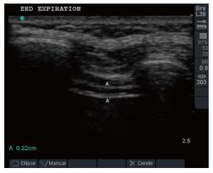
Figure 1 Ultrasonography image of a patient taken at end expiration.
Diaphragm identified as the last set of parallel line, pleural and peritoneal membranes overlying the less echogenic muscle.
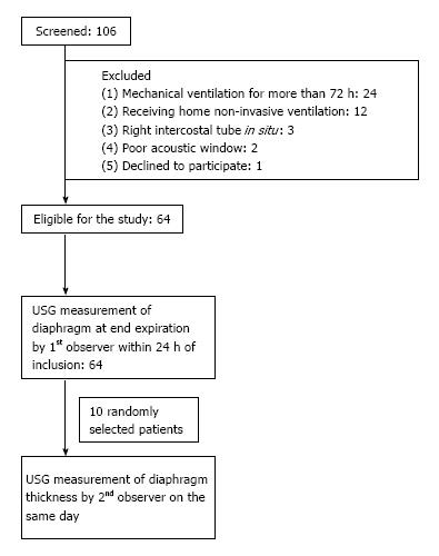
Figure 2 Flow of the patient enrolled into the study.
USG: Ultrasonography.
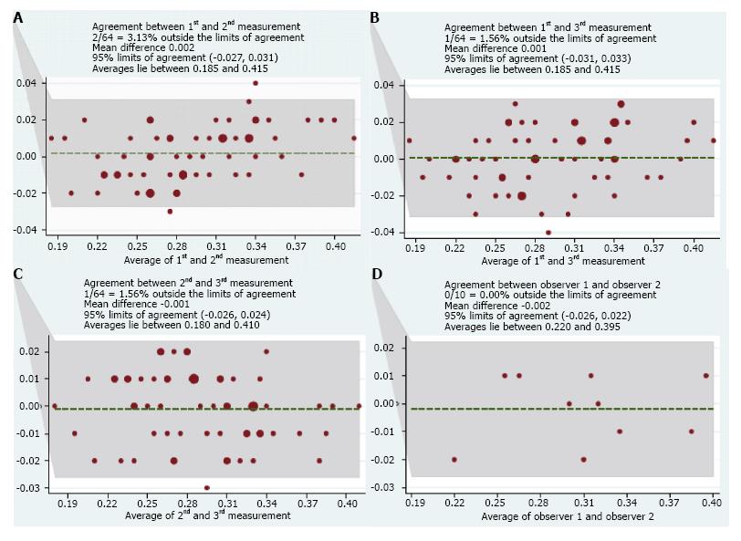
Figure 3 Bland and Altman plots of intraobserver agreement in diaphragm measurement.
The result of three occasions (A-C) and between two observers (D).
- Citation: Dhungana A, Khilnani G, Hadda V, Guleria R. Reproducibility of diaphragm thickness measurements by ultrasonography in patients on mechanical ventilation. World J Crit Care Med 2017; 6(4): 185-189
- URL: https://www.wjgnet.com/2220-3141/full/v6/i4/185.htm
- DOI: https://dx.doi.org/10.5492/wjccm.v6.i4.185


