Copyright
©2014 Baishideng Publishing Group Co.
World J Crit Care Med. Feb 4, 2014; 3(1): 1-7
Published online Feb 4, 2014. doi: 10.5492/wjccm.v3.i1.1
Published online Feb 4, 2014. doi: 10.5492/wjccm.v3.i1.1
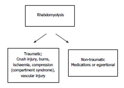
Figure 1 General classification of rhabdomyolysis.
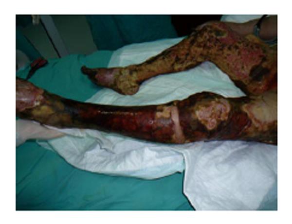
Figure 2 An example for full-thickness burns of both lower extremities.
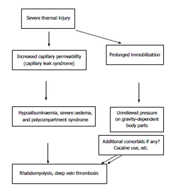
Figure 3 Pathogenic mechanisms for development of rhabdomyolysis in severe thermal injury.
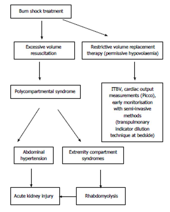
Figure 4 Possible scenarios during burn shock resuscitation.
ITBV: Intrathoracic blood volume.
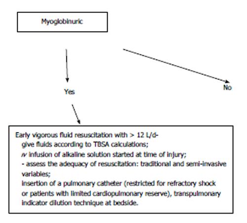
Figure 5 Algorithm for established clinical picture of rhabdomyolysis in burn victims.
TBSA: Total body surface area.
- Citation: Coban YK. Rhabdomyolysis, compartment syndrome and thermal injury. World J Crit Care Med 2014; 3(1): 1-7
- URL: https://www.wjgnet.com/2220-3141/full/v3/i1/1.htm
- DOI: https://dx.doi.org/10.5492/wjccm.v3.i1.1


