Copyright
©The Author(s) 2025.
World J Crit Care Med. Mar 9, 2025; 14(1): 97443
Published online Mar 9, 2025. doi: 10.5492/wjccm.v14.i1.97443
Published online Mar 9, 2025. doi: 10.5492/wjccm.v14.i1.97443
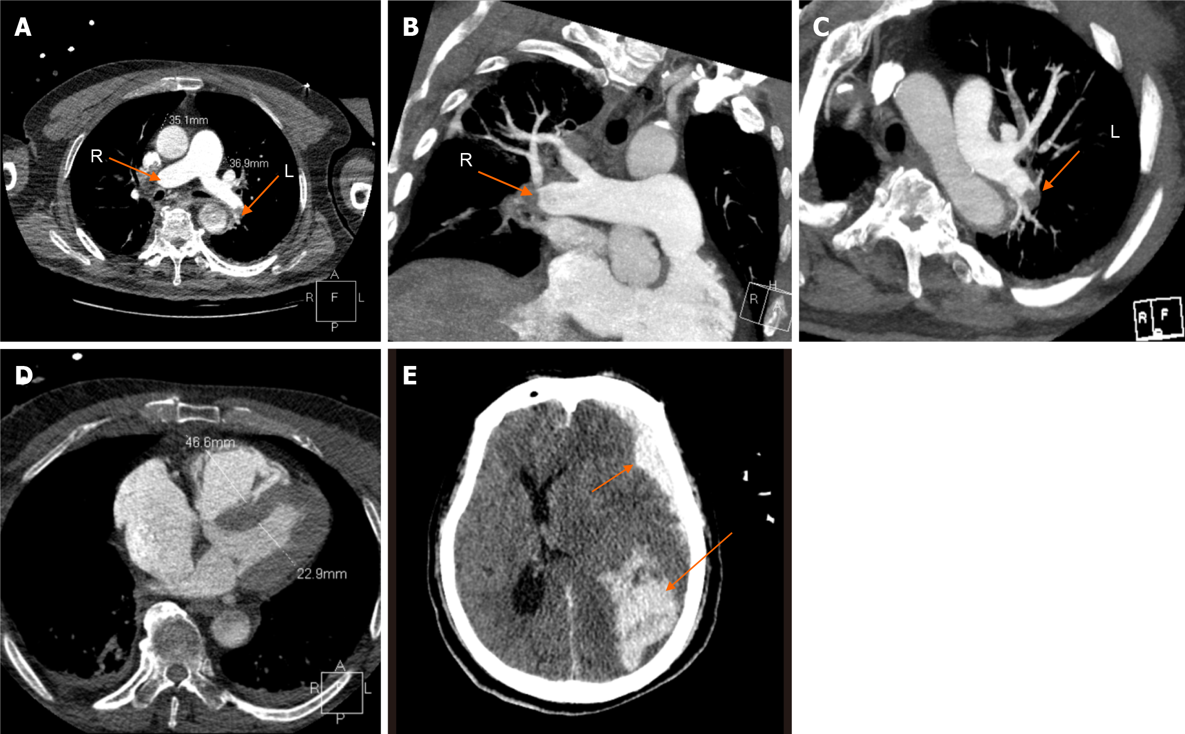
Figure 1 Contrast-enhanced computed tomography findings.
A: Thrombus in both pulmonary arteries (orange arrows); B: Extension to the distal right interlobar artery (orange arrow); C: Extension to the left interlobar pulmonary artery (orange arrow); D: Right ventricle dilation with a diameter of 46.6mm; E: Computed tomography showing left frontal lobe, parietal lobe and subarachnoid hemorrhage (orange arrows).
- Citation: Yuan GX, Zhang ZP, Zhou J. Thrombolysis and extracorporeal cardiopulmonary resuscitation for cardiac arrest due to pulmonary embolism: A case report. World J Crit Care Med 2025; 14(1): 97443
- URL: https://www.wjgnet.com/2220-3141/full/v14/i1/97443.htm
- DOI: https://dx.doi.org/10.5492/wjccm.v14.i1.97443


