Copyright
©The Author(s) 2024.
World J Crit Care Med. Mar 9, 2024; 13(1): 87459
Published online Mar 9, 2024. doi: 10.5492/wjccm.v13.i1.87459
Published online Mar 9, 2024. doi: 10.5492/wjccm.v13.i1.87459
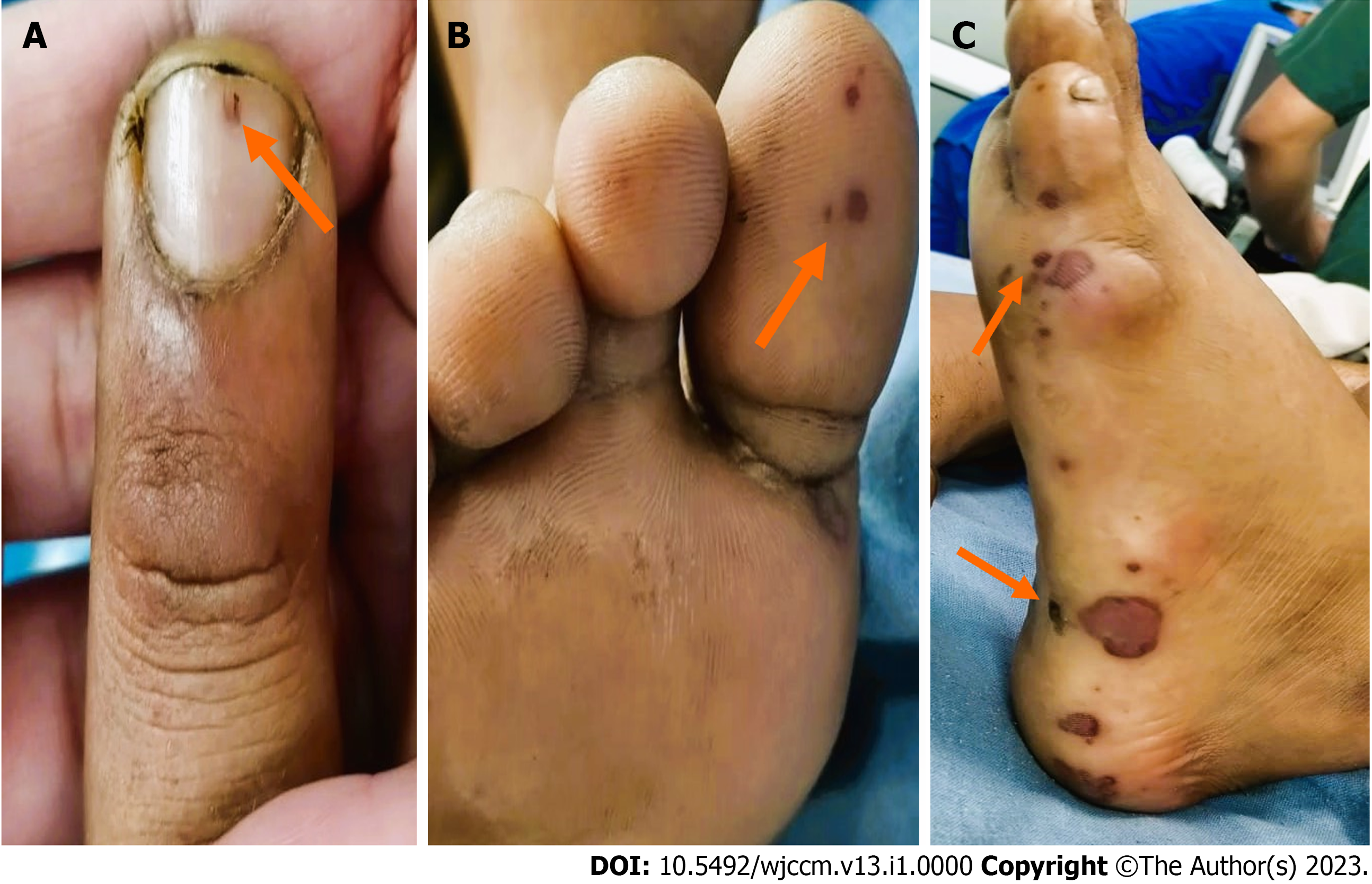
Figure 1 Peripheral manifestations of infective endocarditis.
A: Splinter haemorrhage (arrow) - a minute petechiae on the bed of a fingernail; B and C: Janeway lesions (arrows), multiple small haemorrhages with slight nodularity on the sole of the feet.
Figure 2 Radiological images of the thorax.
A: Frontal X-ray in a sitting position shows bilateral opacities (arrow); B: Computed tomography image through the middle and lower lobes show bilateral lung areas of consolidation and air bronchograms (arrow), along with interspersed areas of ground glass opacities and bilateral pleural effusions (arrow); C: Mediastinal window of the thorax showing cardiomegaly (arrow).
- Citation: Jatteppanavar B, Choudhury A, Panda PK, Bairwa M. Community-acquired multidrug-resistant pneumonia, bacteraemia, and infective endocarditis: A case report. World J Crit Care Med 2024; 13(1): 87459
- URL: https://www.wjgnet.com/2220-3141/full/v13/i1/87459.htm
- DOI: https://dx.doi.org/10.5492/wjccm.v13.i1.87459


