Copyright
©The Author(s) 2022.
World J Crit Care Med. Mar 9, 2022; 11(2): 70-84
Published online Mar 9, 2022. doi: 10.5492/wjccm.v11.i2.70
Published online Mar 9, 2022. doi: 10.5492/wjccm.v11.i2.70
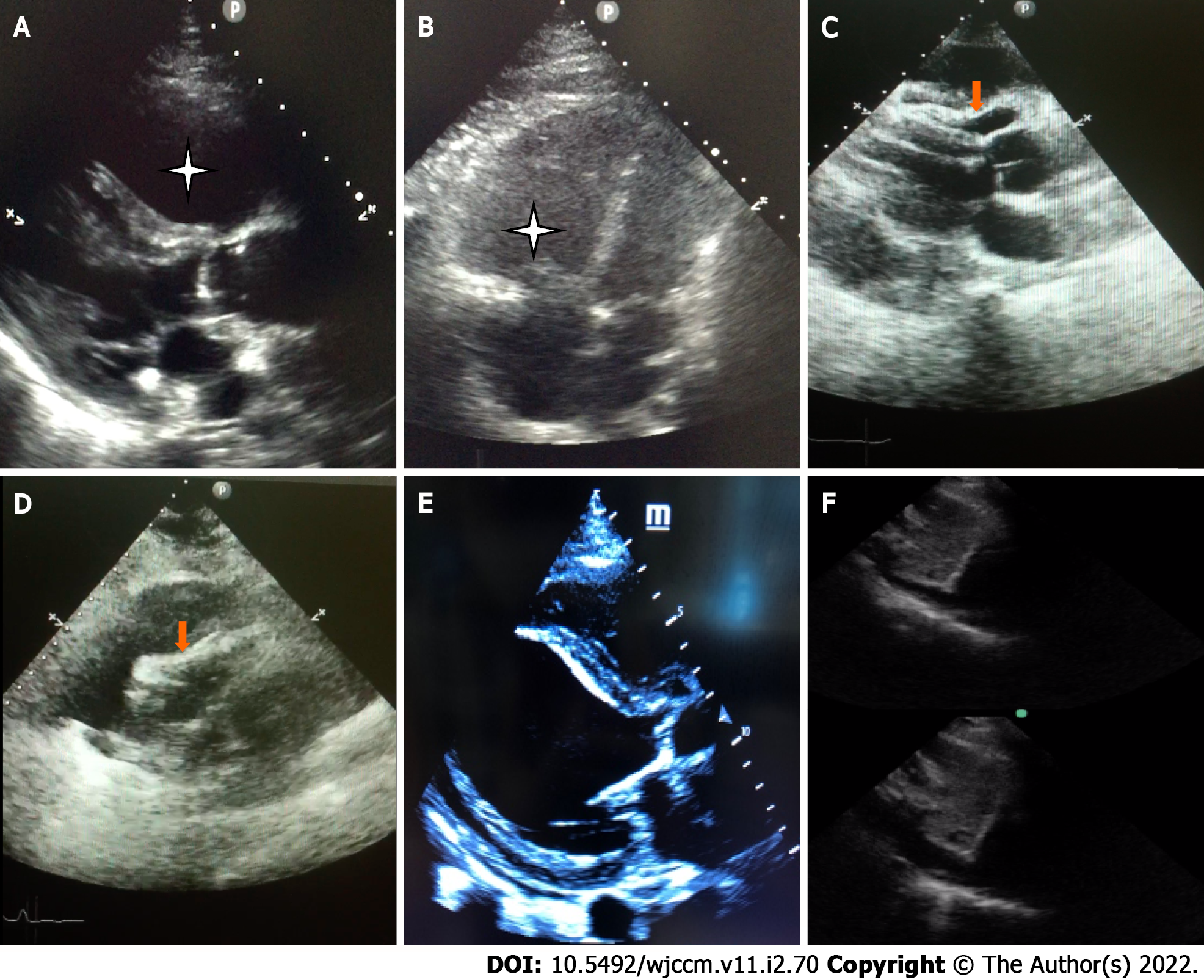
Figure 1 Key features in basic critical care echocardiography.
A: Dilated right ventricle [Parasternal long axis (PLAX)]; B: Dilated right ventricle (Apical 4 chamber view); C: Pericardial tamponade-Pericardial effusion with diastolic collapse of right ventricle (PLAX view); D: Pericardial tamponade–Pericardial effusion with systolic collapse of right atrium [subcostal long axis (SLAX) view]; E: Left ventricular dysfunction-minimal thickening and contraction of basal anteroseptal and inferolateral wall with severe hypokinesia (PLAX view); F: Inferior vena cava variation of > 50% with foreceful spontaneous respiration-“sniff test” (SLAX view).
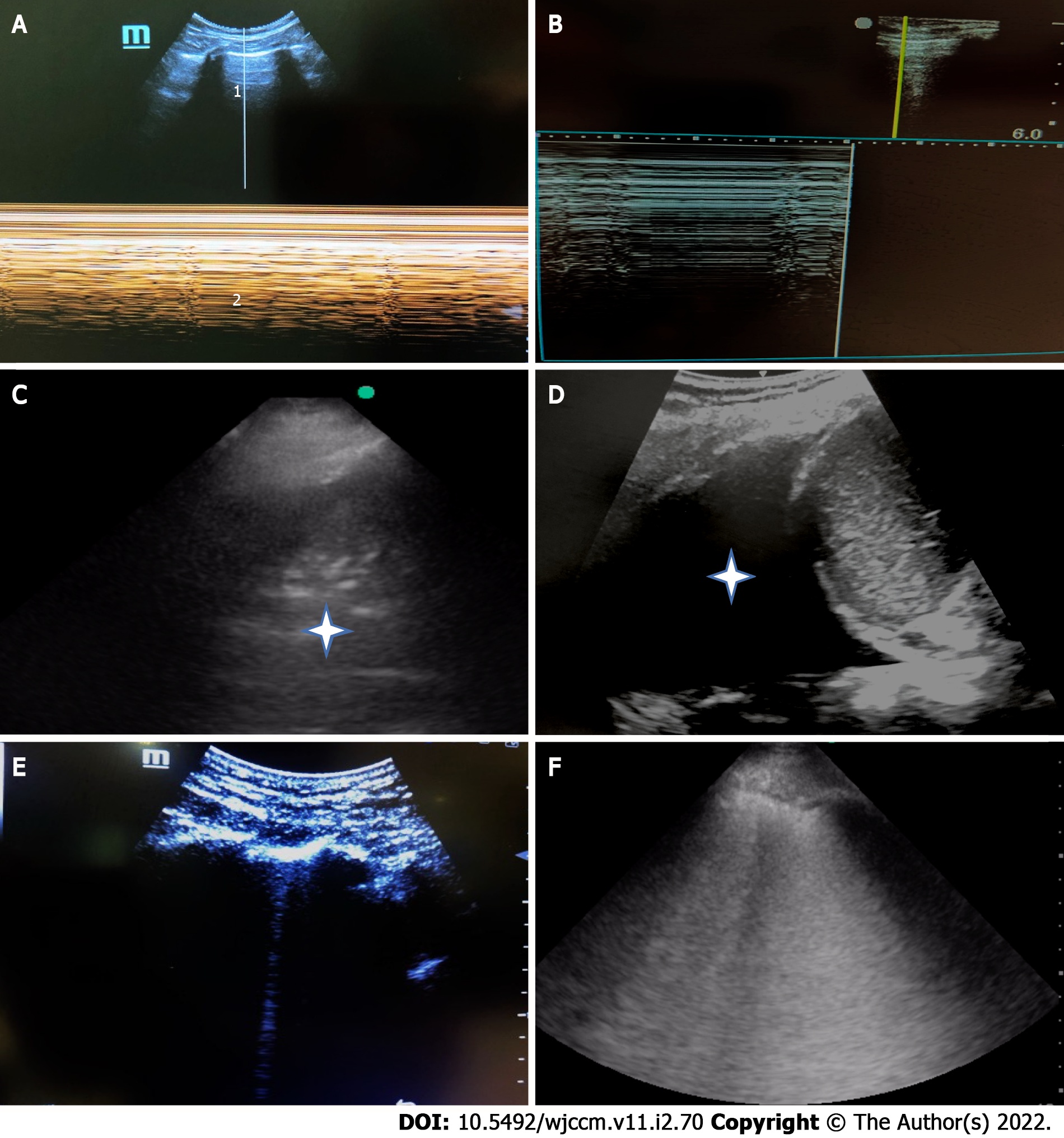
Figure 2 Key features in basic lung ultrasound.
A: M-mode lung ultrasound-normal a lines (1), and seashore sign (2); B: M-mode lung ultrasound-pneumothorax Bar code/stratosphere sign; C: Consolidation with air bronchograms (Asterisk); D: Pleural effusion (large); E: 1 single B line-normal; F: B profile, > 3 B lines (confluent)-pathological.
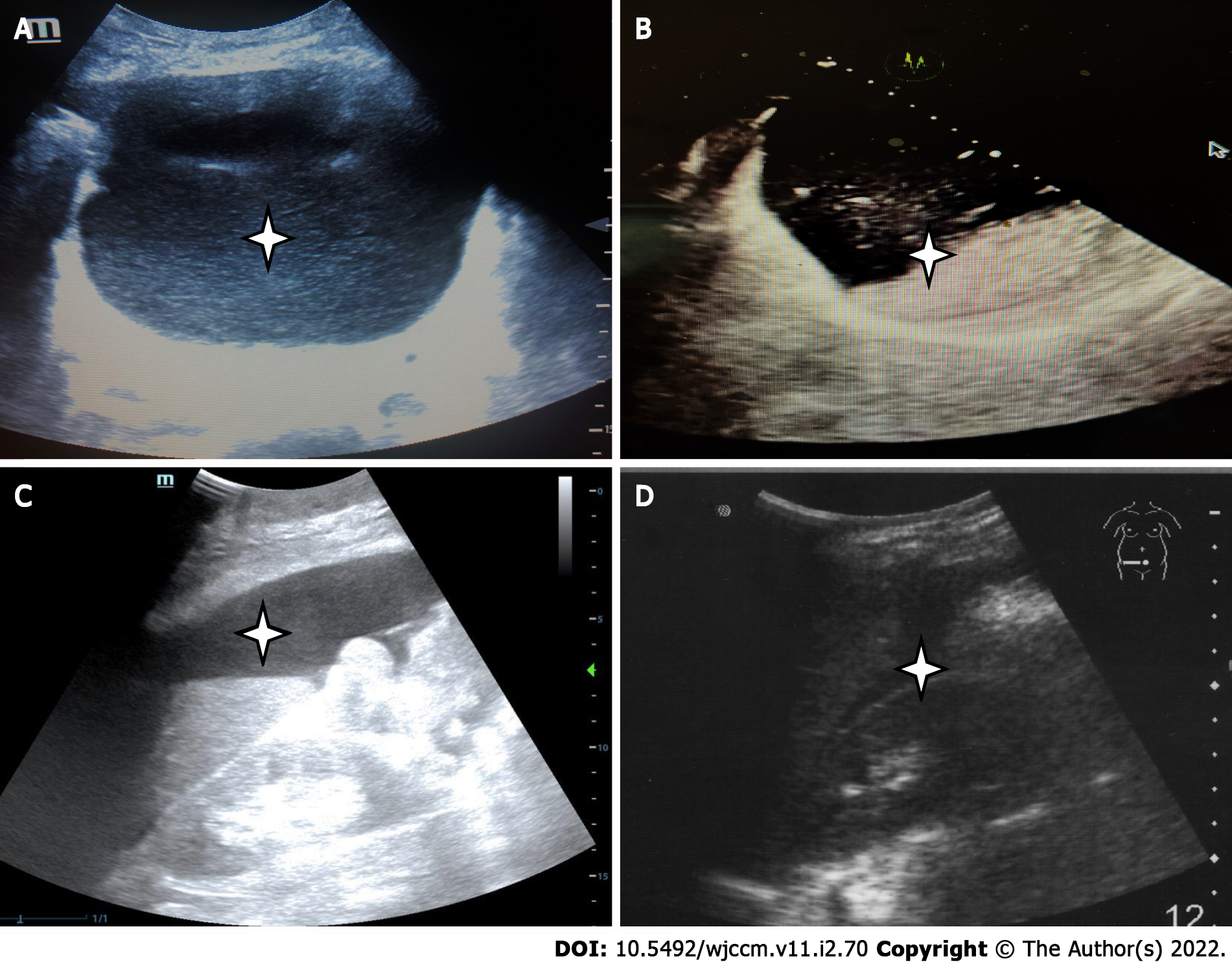
Figure 3 Key features in abdominal ultrasound.
A: Bladder overdistension due to acute retention of urine (Asterisk); B: Incomplete gastric emptying (presence of semi-digested food in the stomach, Asterisk), which will indicate need for rapid sequence induction for intubation; C: Ascites (Asterisk); D: Free fluid in the hepato-renal pouch. In cases with abdominal trauma, this indicates intra-peritoneal bleeding (Asterisk).
- Citation: Lau YH, See KC. Point-of-care ultrasound for critically-ill patients: A mini-review of key diagnostic features and protocols. World J Crit Care Med 2022; 11(2): 70-84
- URL: https://www.wjgnet.com/2220-3141/full/v11/i2/70.htm
- DOI: https://dx.doi.org/10.5492/wjccm.v11.i2.70


