Copyright
©The Author(s) 2021.
World J Crit Care Med. Sep 9, 2021; 10(5): 301-309
Published online Sep 9, 2021. doi: 10.5492/wjccm.v10.i5.301
Published online Sep 9, 2021. doi: 10.5492/wjccm.v10.i5.301
Figure 1 Chest radiograph.
A: Plain AP erect chest radiograph taken shortly after arrival to the emergency room, showing dense consolidation of right middle and lower zones; B: Plain AP erect chest radiograph following tracheal intubation and central line insertion. Note progression of consolidation as seen in Figure 1A and changes in keeping with acute respiratory distress syndrome; C: Plain chest radiograph taken approximately 8 h after presentation. Note dense consolidation and air bronchograms seen in right lower lobe, progressing from previous chest radiographs.
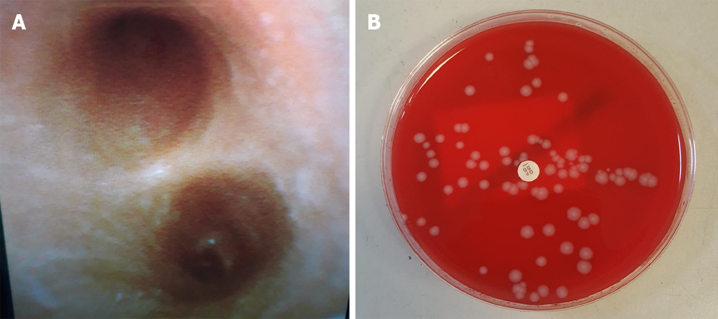
Figure 2 Emergency therapeutic and diagnostic bronchoscopy was performed in Adult Intensive Care Units.
A: Bronchoscopic appearances of carina. Samples taken with bronchoalveolar lavage confirmed as Panton-Valentine leukocidin (PVL)-Staphylococcus aureus (S. aureus) and influenza A/H3N2; B: Blood agar plate showing colony forming units of S. aureus from bronchial alveolar lavage performed in Adult Intensive Care Units, subsequently identified as PVL producing S. aureus from Public Health England reference laboratory using whole genome sequencing.
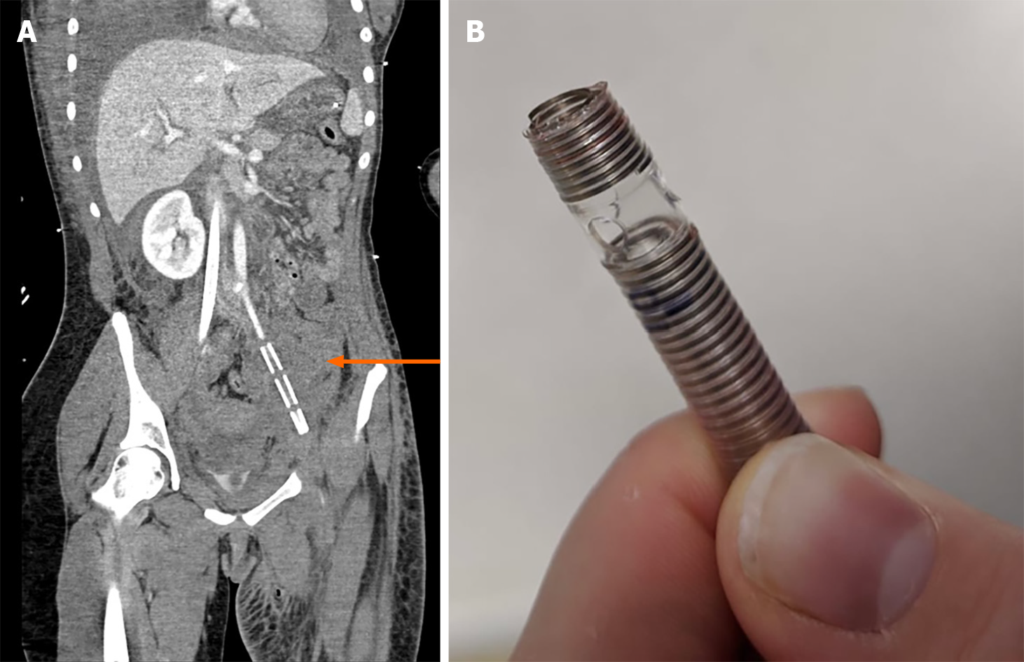
Figure 3 Computed tomography.
A: Coronal computed tomography scan slice demonstrating retained extracorporeal membrane oxygenation (ECMO) cannula fragment (orange arrow); B: The torn end of the partially removed ECMO cannula.
Figure 4 Timeline of key events with time since initial presentation.
ICU: Intensive care units; ECMO: Extracorporeal membrane oxygenation.
- Citation: Cuddihy J, Patel S, Mughal N, Lockie C, Trimlett R, Ledot S, Cheshire N, Desai A, Singh S. Near-fatal Panton-Valentine leukocidin-positive Staphylococcus aureus pneumonia, shock and complicated extracorporeal membrane oxygenation cannulation: A case report. World J Crit Care Med 2021; 10(5): 301-309
- URL: https://www.wjgnet.com/2220-3141/full/v10/i5/301.htm
- DOI: https://dx.doi.org/10.5492/wjccm.v10.i5.301


