Copyright
©2012 Baishideng.
World J Crit Care Med. Aug 4, 2012; 1(4): 102-105
Published online Aug 4, 2012. doi: 10.5492/wjccm.v1.i4.102
Published online Aug 4, 2012. doi: 10.5492/wjccm.v1.i4.102
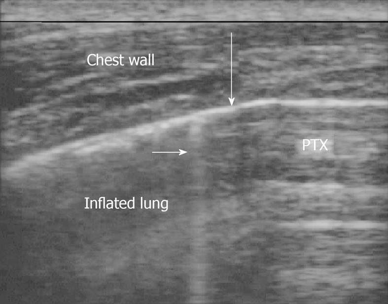
Figure 1 An ultrasound depiction of a pneumothorax is shown.
This is the lung point. To the right of the image air blocks visualization of typical lung artifacts. On the left, the visceral and parietal pleura are sliding past each other. The large arrow shows where they meet. The small arrow shows a B line, seen only in inflated portions of the lung.
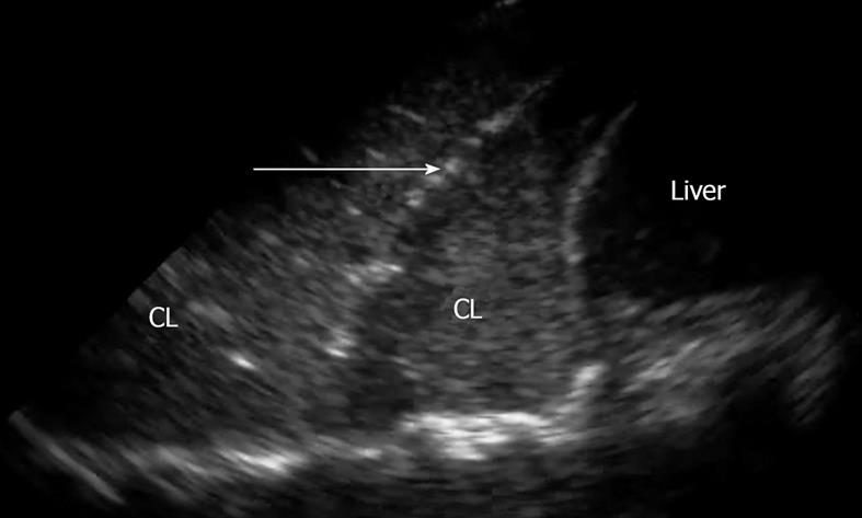
Figure 2 Pneumonia is seen just above the liver and diaphragm.
An air bronchogram is also seen (arrow). Air can actually be seen moving through the bronchus within the consolidated, infected lung in real time. CL: Consolidated lung.
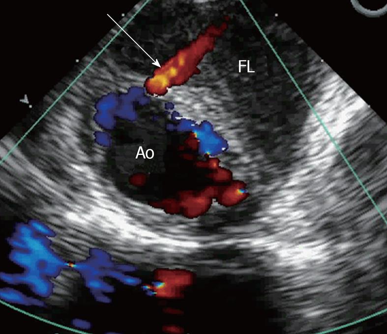
Figure 3 A jet of blood (arrow) is seen dissecting from the true lumen into the false lumen in this patient with a thoracic aortic dissection.
Ao: Aorta; FL: False lumen.
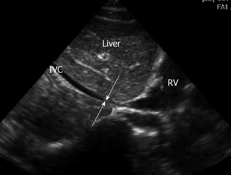
Figure 4 Inferior vena cava flat in this hypotensive and volume depleted patient.
Arrows show the barely open inferior vena cava suggesting need to considerable fluid resuscitation to correct hypotension. RV: Right ventricle of the heart; IVC: Inferior vena cava.
- Citation: Blaivas M. Update on point of care ultrasound in the care of the critically ill patient. World J Crit Care Med 2012; 1(4): 102-105
- URL: https://www.wjgnet.com/2220-3141/full/v1/i4/102.htm
- DOI: https://dx.doi.org/10.5492/wjccm.v1.i4.102


