Copyright
©The Author(s) 2015.
World J Surg Proced. Mar 28, 2015; 5(1): 82-98
Published online Mar 28, 2015. doi: 10.5412/wjsp.v5.i1.82
Published online Mar 28, 2015. doi: 10.5412/wjsp.v5.i1.82
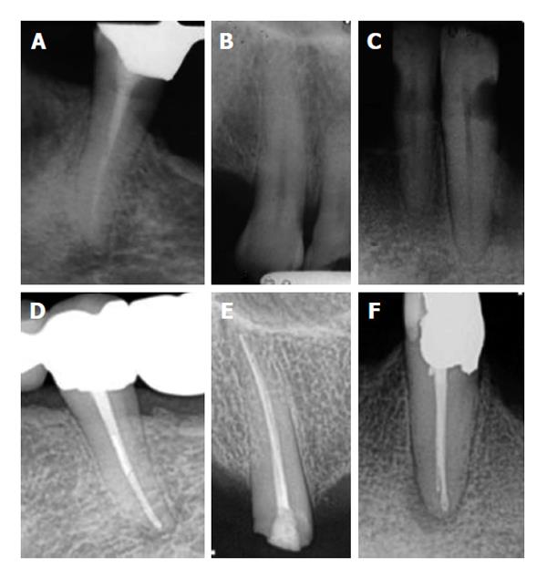
Figure 1 (A-C) preoperative and (D-E) 5-year follow-up radiographs: (D) complete healing, (E) uncertain healing, and (F) no healing.
Figure 2 The extractor is inserted into the groove and locked the end of the fragment by the screw tightened between the plunger and the internal embossment.
A: Periapical radiograph: Separated instrument is visible in middle 3rd of calcified root canal in maxillary right lateral incisor; B and C: Making a channel around the separated instrument to keep the broken instrument in the center of the tube of Masserann Kit; D and E: Engaging tube of Masserann Kit with the separated instrument and removal of the fragment from root canal.
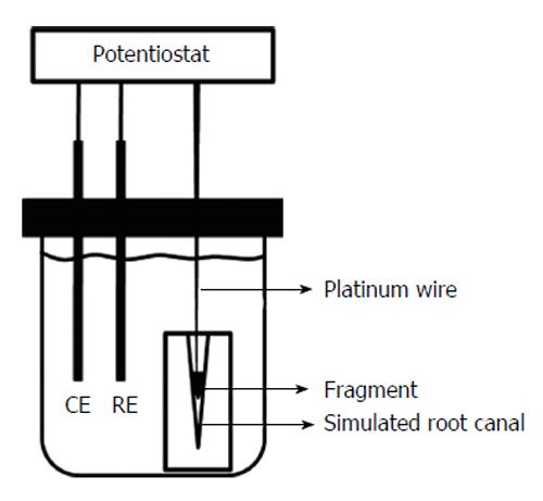
Figure 3 Schematic diagram of the intracanal fragment dissolution test.
CE: Counter electrode; RE: Reference electrode.
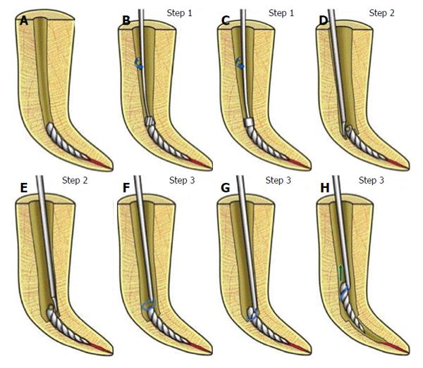
Figure 4 Procedures for removing a separated file from a root canal using the new file removal system.
A: Initial canal with a separated file; B: Canal enlarged with CBA; C: Dentin removal around the separated file with CBB; D: Ultrasonic tip troughed semicircularly around the separated file to create space for the file-removal device; E: troughing semicircularly on the remaining half of the separated file for complete exposure; F: Placement of the loop over the separated file; G: Fastening the loop to grab the separated file; H: Removal of the separated file from the root canal.
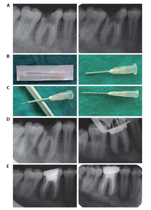
Figure 5 A modified 18-gauge needle and cyanoacrylate glue were used to retrieve a separated nickel-titanium instrument from the mesiolingual canal of a mandibular first molar.
A: Radiograph showing separated instrument; Radiograph showing dentine surrounding the coronalend of the separated fragment removed with GG drill; B: An 18-guage needle, modified by cutting with a carborundum disc from the tip to transform it into a microtube; C: Separated instrument fragment removed adhered to the microtube; D: Radiograph confirming instrument removal; Working length reconfirmed; Post-obturation radiograph; E: Two-year follow up radiograph.
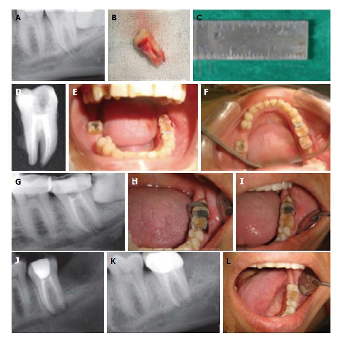
Figure 6 A separated hand instrument in a second molar was retrieved from the mesiobuccal root, which was close to the mandibular canal, using tooth replantation.
A: Broken instrument near to the mandibular canal; B: After extraction; C: Measured broken instrument of 7 mm; D: After obturation; E: After separators were placed; F: Extra coronal splinting with orthodontic wires were prepared; G: Post operative Radiograph; H and I: Four weeks after Band removal; J: Three months follow-up Radiograph; K: One year follow-up radiograph; L: One year clinical radiograph.
- Citation: Tang WR, Smales RJ, Chen HF, Guo XY, Si HY, Gao LM, Zhou WB, Wu YN. Prevention and management of fractured instruments in endodontic treatment. World J Surg Proced 2015; 5(1): 82-98
- URL: https://www.wjgnet.com/2219-2832/full/v5/i1/82.htm
- DOI: https://dx.doi.org/10.5412/wjsp.v5.i1.82


