Copyright
©The Author(s) 2015.
World J Surg Proced. Mar 28, 2015; 5(1): 111-118
Published online Mar 28, 2015. doi: 10.5412/wjsp.v5.i1.111
Published online Mar 28, 2015. doi: 10.5412/wjsp.v5.i1.111
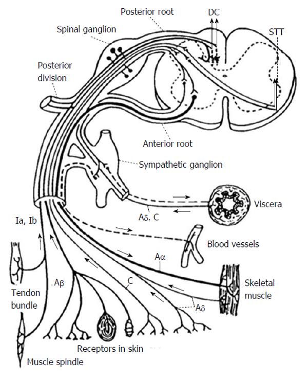
Figure 1 Peripheral and central spinal pathways.
Aδ: Large myelinated afferent fiber. Type A fibers don’t conduct nociception. C fibers are small unmyelinated fibers that conduct nociception. Ia and Ib fibers conduct information from muscle spindles and Golgi tendon organs, respectively. DC: Dorsal columns; STT: Spinothalamic tract.
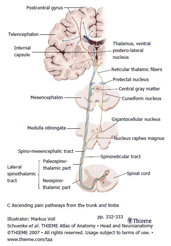
Figure 2 The relationship between ascending pain afferents and their targets in the medulla, midbrain, thalamus and cortex.
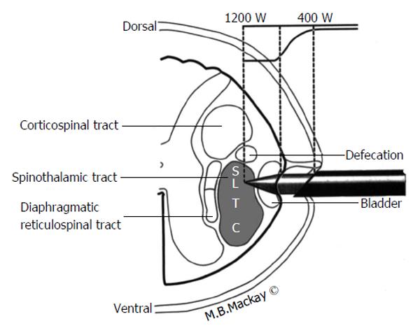
Figure 3 A schematic illustration of percutaneous cordotomy and the relative anatomical position of the spinothalamic tract and surrounding structures.
S: Sacral; L: Lumbar; T: Thoracic; C: Cervical; W: Watts.
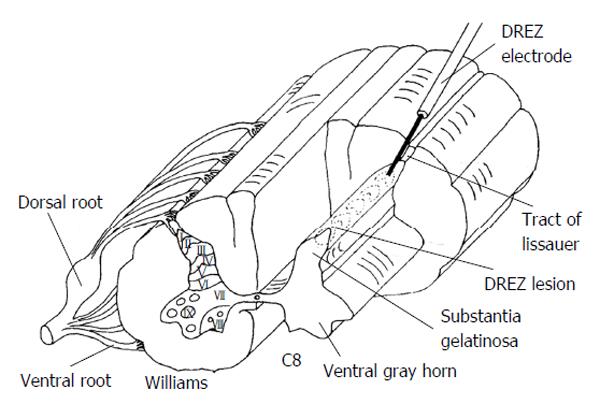
Figure 4 A schematic illustrating dorsal root entry zone lesioning at the C8 spinal level.
Rexed lamina are numbered in the typical fashion I-XIII. DREZ: Dorsal root entry zone.
- Citation: Lake WB, Konrad PE. Cordotomy procedures for cancer pain: A discussion of surgical procedures and a review of the literature. World J Surg Proced 2015; 5(1): 111-118
- URL: https://www.wjgnet.com/2219-2832/full/v5/i1/111.htm
- DOI: https://dx.doi.org/10.5412/wjsp.v5.i1.111


