Copyright
©2013 Baishideng Publishing Group Co.
World J Surg Proced. Nov 28, 2013; 3(3): 41-46
Published online Nov 28, 2013. doi: 10.5412/wjsp.v3.i3.41
Published online Nov 28, 2013. doi: 10.5412/wjsp.v3.i3.41
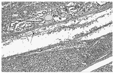
Figure 1 Hematoxilin-Eosin staining shows micropapillary carcinoma (diameter 10 mm, arrow, upper part) with follicular adenoma (diameter 50 mm, lower part).
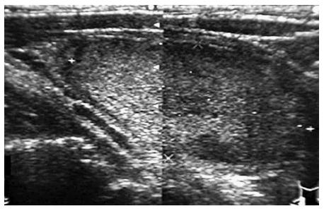
Figure 2 A tumor diameter of approximately 60 mm, solid and accounts for most of the left lobe were suggested by ultrasound.
Although capsular invasion was unclear, Doppler examination were revealed abundant blood flow and high blood flow resistance value inside the tumor. The result of ultrasound suspected the malignancy.
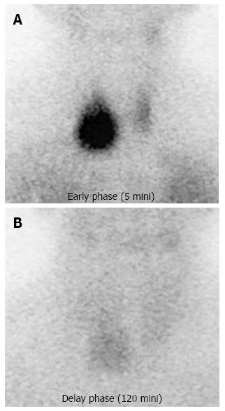
Figure 3 In the Tl early phase of 5 min after injection, a prominent accumulation of isotope to the tumor was observed.
In the delay phase of 120 min after injection, a remaining of isotope in tumor was also found. These accumulations of isotope and delay of wash out suggest the suspicion of malignancy findings.
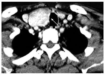
Figure 4 Computed tomography image of follicular neoplasm of 45 mm diameter in the right lobe.
It showed some irregular border and extension of the right wall of the trachea. There is a suspicion of tracheal invasion of tumor.
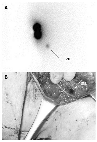
Figure 5 Sentinel nodes.
A: Sentinel nodes (SNL, arrow) revealed in the central component lymph nodes using Tc-physic acid. B: SNL (arrow) is performed using 4% isosulfan blue dye during operation. The stained lymph node is seen near the right pharyngeal recurrent nerve in the central component lymph nodes.
- Citation: Takeyama H, Tabei I, Kato K, Kamio M, Nogi H, Toriumi Y, Kinoshita S, Akiba T, Uchida K, Morikawa T. Operative indications of follicular type tumors, based on Japanese clinical guidelines. World J Surg Proced 2013; 3(3): 41-46
- URL: https://www.wjgnet.com/2219-2832/full/v3/i3/41.htm
- DOI: https://dx.doi.org/10.5412/wjsp.v3.i3.41


