Copyright
©The Author(s) 2024.
World J Surg Proced. Mar 1, 2024; 14(2): 8-14
Published online Mar 1, 2024. doi: 10.5412/wjsp.v14.i2.8
Published online Mar 1, 2024. doi: 10.5412/wjsp.v14.i2.8
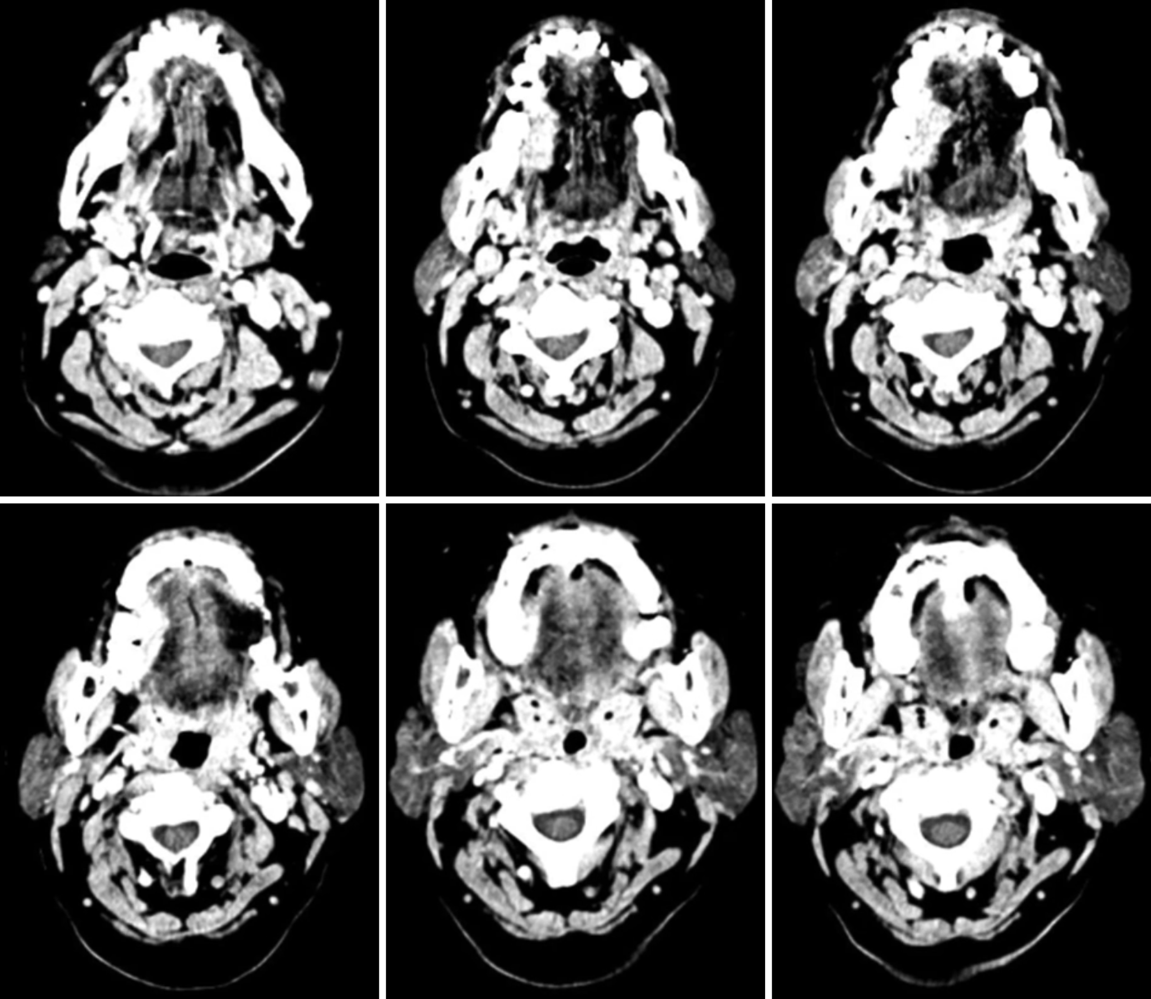
Figure 1 Multi-slice computed tomography of the midface.
Mass of the right tongue with contrast enhancement.
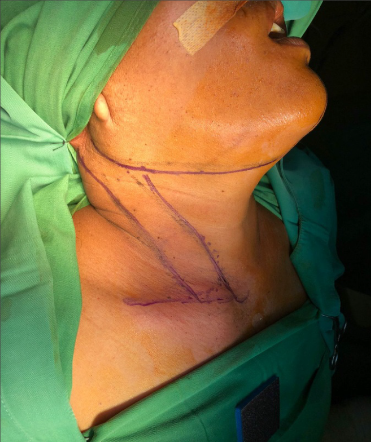
Figure 2 Surgical site marking.
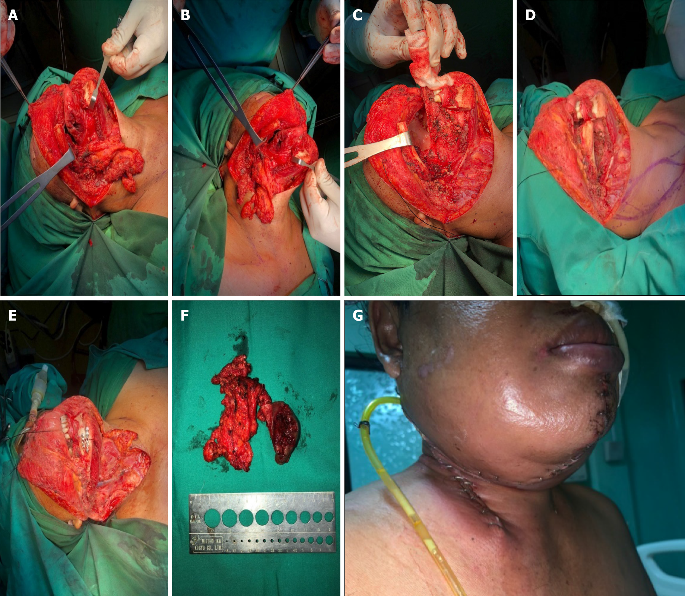
Figure 3 Intraoperative procedure.
A-C: Wide excision right tongue with neck and mandibular dissection; D: Sternocleidomastoid (SCM) flap design; E: After reconstruction of SCM flap for floor of the mouth and fixation of mandible bone; F: Mass excised from the right tongue, neck, and mandible; G: After surgery at ward with drain.
- Citation: Irawan H, Bharata MBS. Sternocleidomastoid flap for reconstruction of tongue small cell carcinoma: A case report. World J Surg Proced 2024; 14(2): 8-14
- URL: https://www.wjgnet.com/2219-2832/full/v14/i2/8.htm
- DOI: https://dx.doi.org/10.5412/wjsp.v14.i2.8


