Copyright
©The Author(s) 2024.
World J Surg Proced. Feb 2, 2024; 14(1): 1-7
Published online Feb 2, 2024. doi: 10.5412/wjsp.v14.i1.1
Published online Feb 2, 2024. doi: 10.5412/wjsp.v14.i1.1
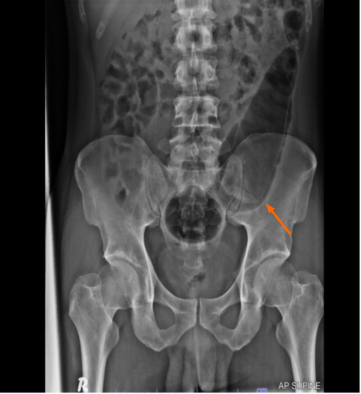
Figure 1
Plain abdominal radiograph showed bottle-shaped foreign body in the left sided of abdomen (orange arrow).
Figure 2 Laparoscopic view of sigmoid colon.
A and B: Following the shape of foreign body. The sigmoid colon serosa was normal looking and non edematous.
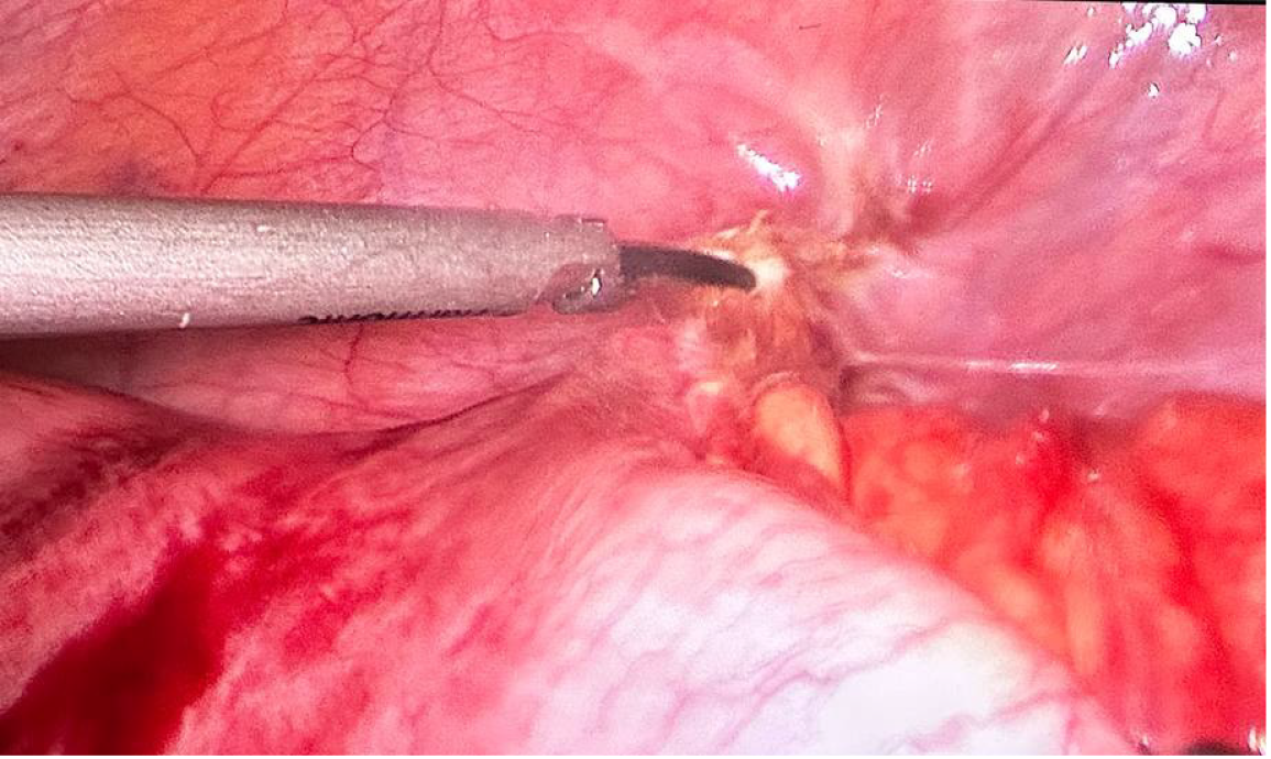
Figure 3
Adhesiolysis performed to release adhesion to straighten the axis.
Figure 4 Surgical diagram.
A and B: A 5-mm laparoscopic atraumatic bowel grasper and babcock forcep were used to help pushed down the foreign body into the rectum, with traction and counter-traction methods.
Figure 5 Surgical diagram.
A and B: The foreign body managed to pass down into the rectum.
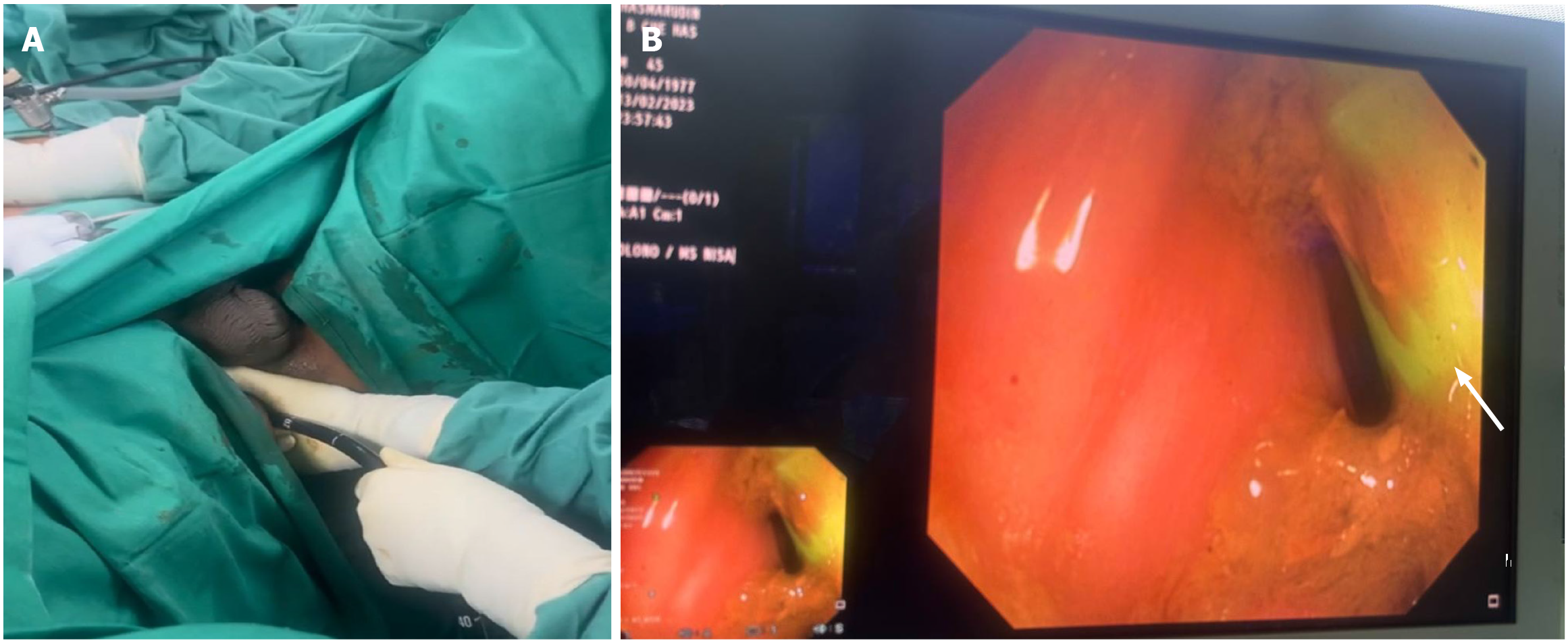
Figure 6 The rectal mucosa was not inflamed nor oedematous.
A: The assistant is doing sigmoidoscopy; B: Endoscopic image of rectum, with greenish foreign body (white arrow).
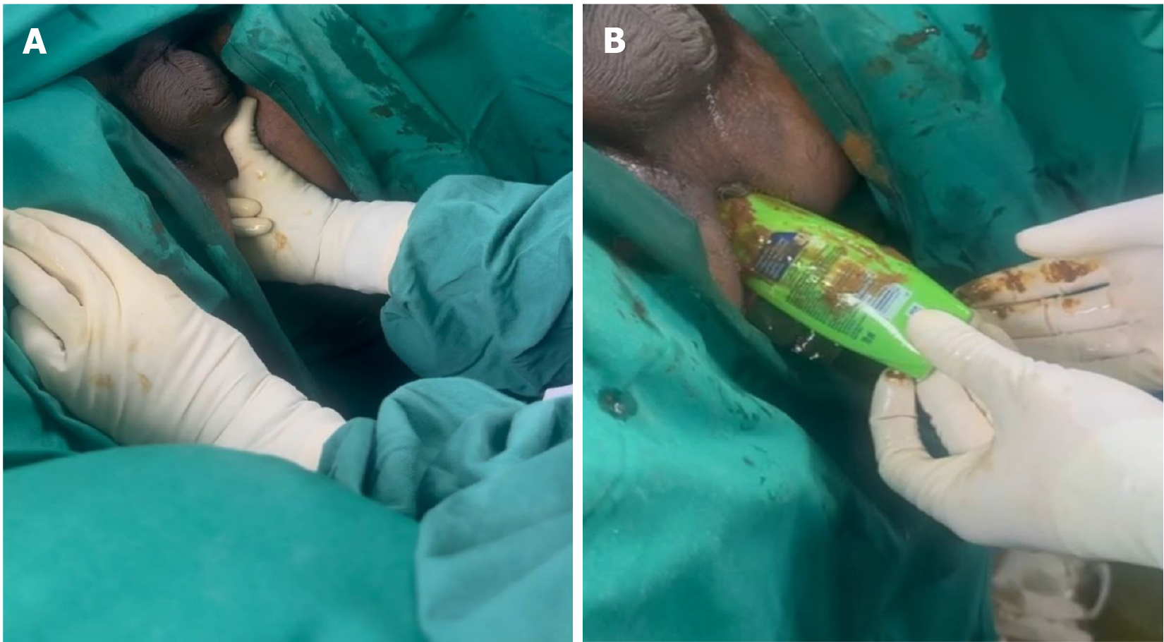
Figure 7 The first surgeon pushed the FB toward the anus via laparoscopy until it reached the second surgeon’s hand in the anus.
A: Bimanual palpation of foreign body via suprapubic and transanally; B: Successful retrieval of the foreign body.
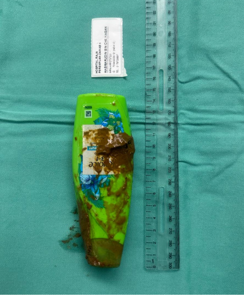
Figure 8
The removed shampoo bottle.
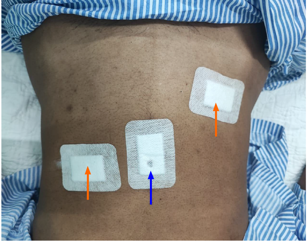
Figure 9 Port placement area.
Blue arrow: 12-mm camera port at infraumbilical. Orange arrow: 5-mm working at right iliac fossa and left lumbar.
- Citation: Che Ghazali K, Yaacob H, Mohamed Sidek AS. Combined laparoscopic and endoscopic method for foreign body removal from descending colon: A case report. World J Surg Proced 2024; 14(1): 1-7
- URL: https://www.wjgnet.com/2219-2832/full/v14/i1/1.htm
- DOI: https://dx.doi.org/10.5412/wjsp.v14.i1.1


