Copyright
©The Author(s) 2023.
World J Surg Proced. Dec 28, 2023; 13(3): 14-21
Published online Dec 28, 2023. doi: 10.5412/wjsp.v13.i3.14
Published online Dec 28, 2023. doi: 10.5412/wjsp.v13.i3.14
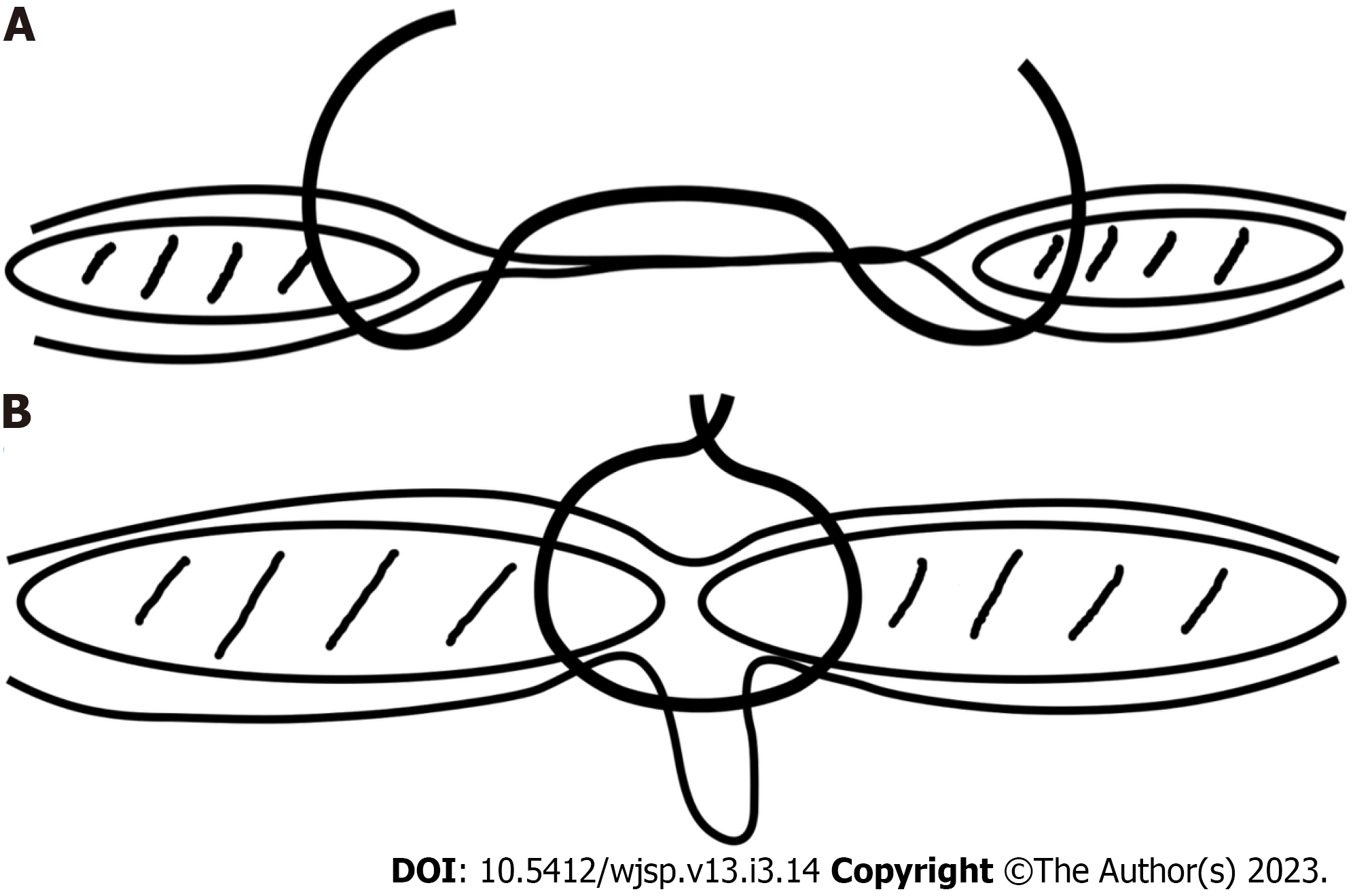
Figure 1 Rectus muscle repair technique.
A: The suture engages full thickness of rectus abdominis muscle and its anterior and posterior sheaths; B: When pulled together, the recti assume a midline position, inverting the attenuated linea alba and hernia sac.
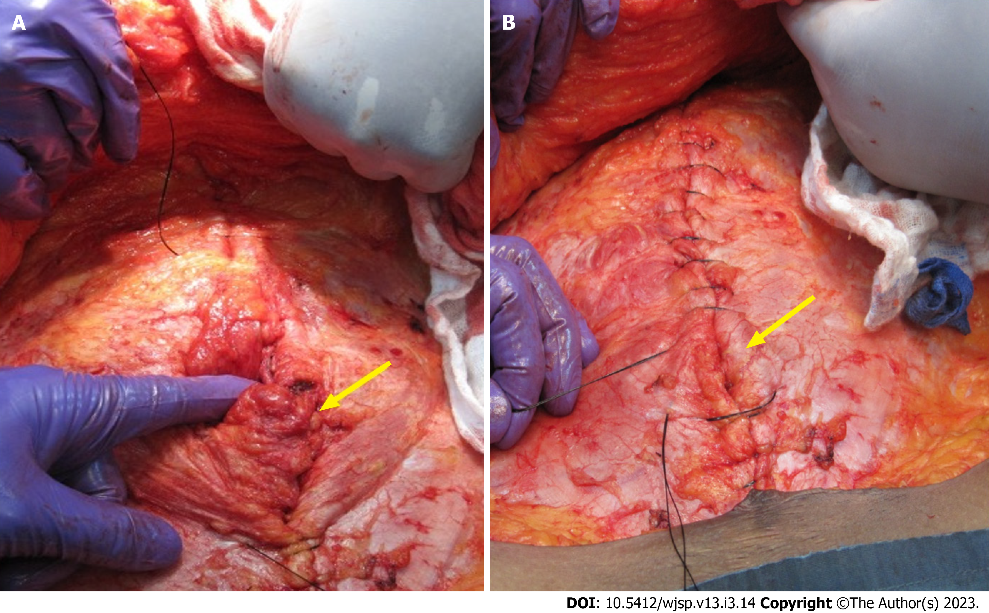
Figure 2 The hernia sac and attenuated linea alba are inverted by placing full-thickness sutures to approximate the rectus abdominis muscles (arrows).
A: The sutures incorporate the anterior and posterior sheaths en masse including the medial 1-1.5 cm of the muscle; B: All attenuated midline tissues are therefore eliminated (by inverting them).
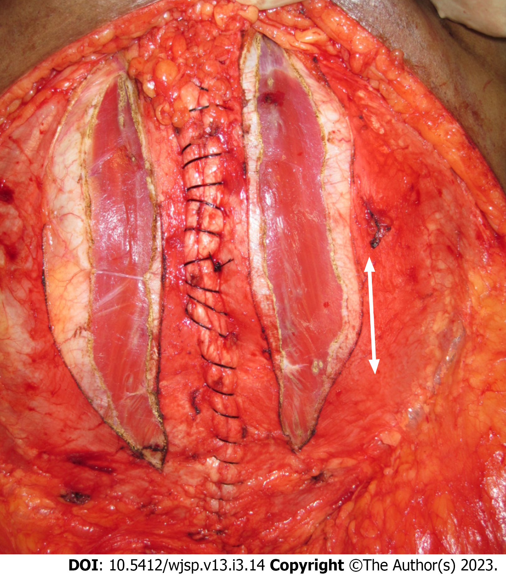
Figure 3 Relaxing incisions are made in the anterior rectus sheath in order to reduce tension on the suture line.
- Citation: Naraynsingh V, Cawich SO, Hassranah S. Alternative to mesh repair for ventral hernias: Modified rectus muscle repair. World J Surg Proced 2023; 13(3): 14-21
- URL: https://www.wjgnet.com/2219-2832/full/v13/i3/14.htm
- DOI: https://dx.doi.org/10.5412/wjsp.v13.i3.14


