Copyright
©The Author(s) 2023.
World J Surg Proced. Sep 28, 2023; 13(2): 7-13
Published online Sep 28, 2023. doi: 10.5412/wjsp.v13.i2.7
Published online Sep 28, 2023. doi: 10.5412/wjsp.v13.i2.7
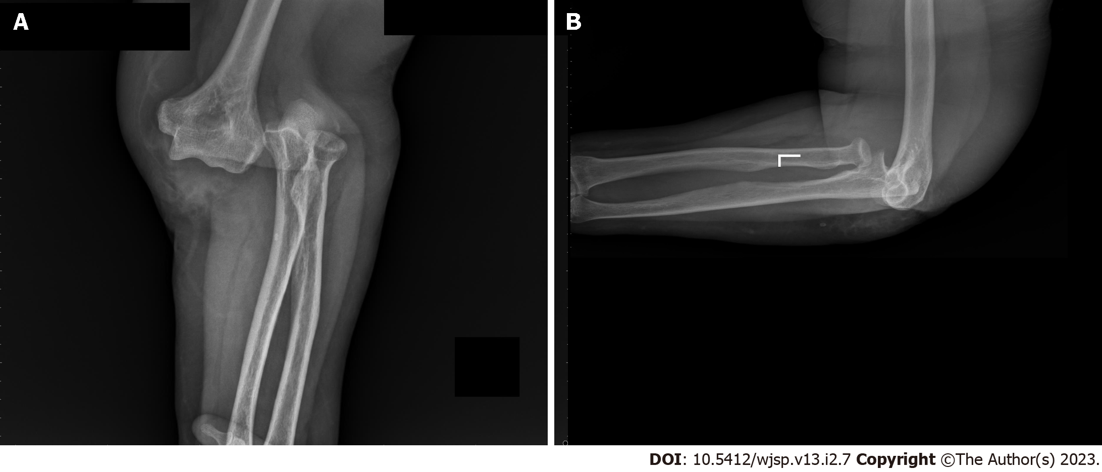
Figure 1 Roentgenographs at the time of the injury.
A: Anteroposterior; B: Lateral.
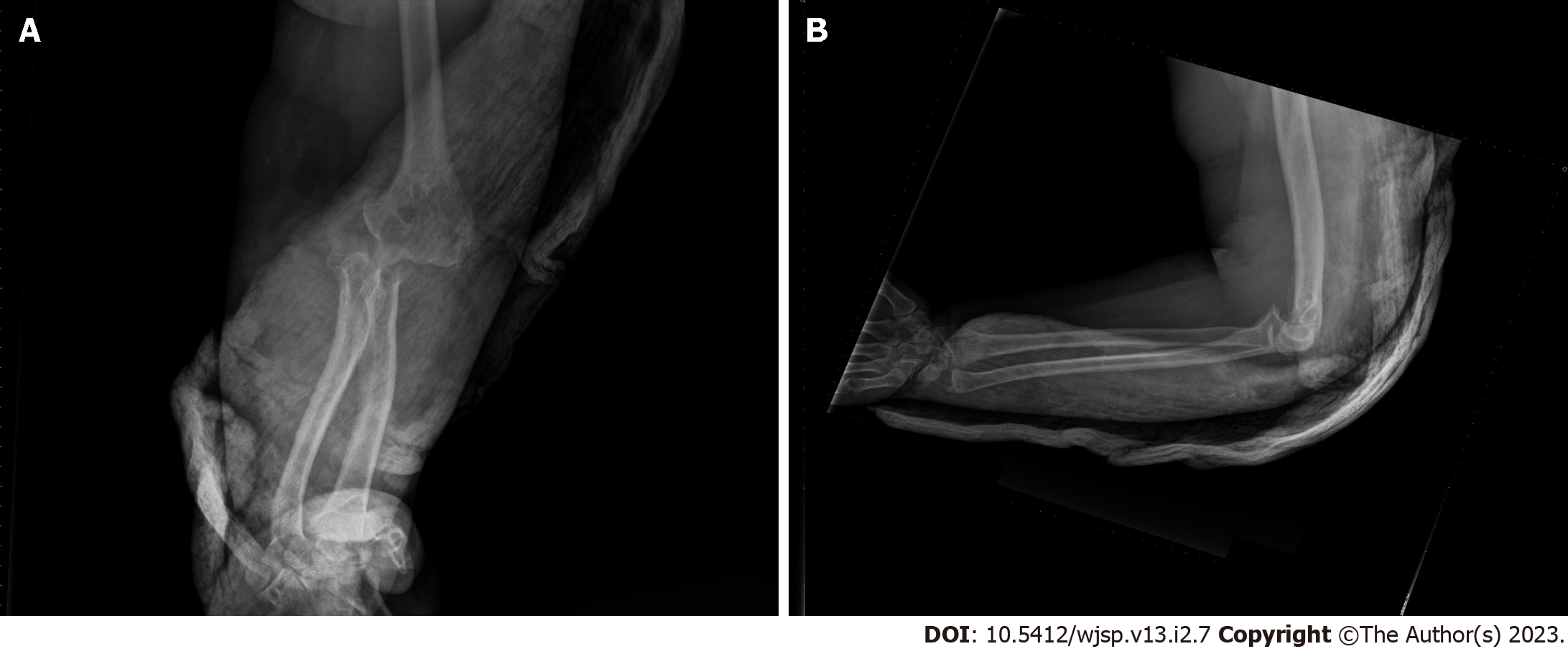
Figure 2 Roentgenographs after closed reduction.
A: Anteroposterior; B: Lateral.
Figure 3 Roentgenographs after one week.
Loss of reduction is seen. A: Anteroposterior; B: Lateral.
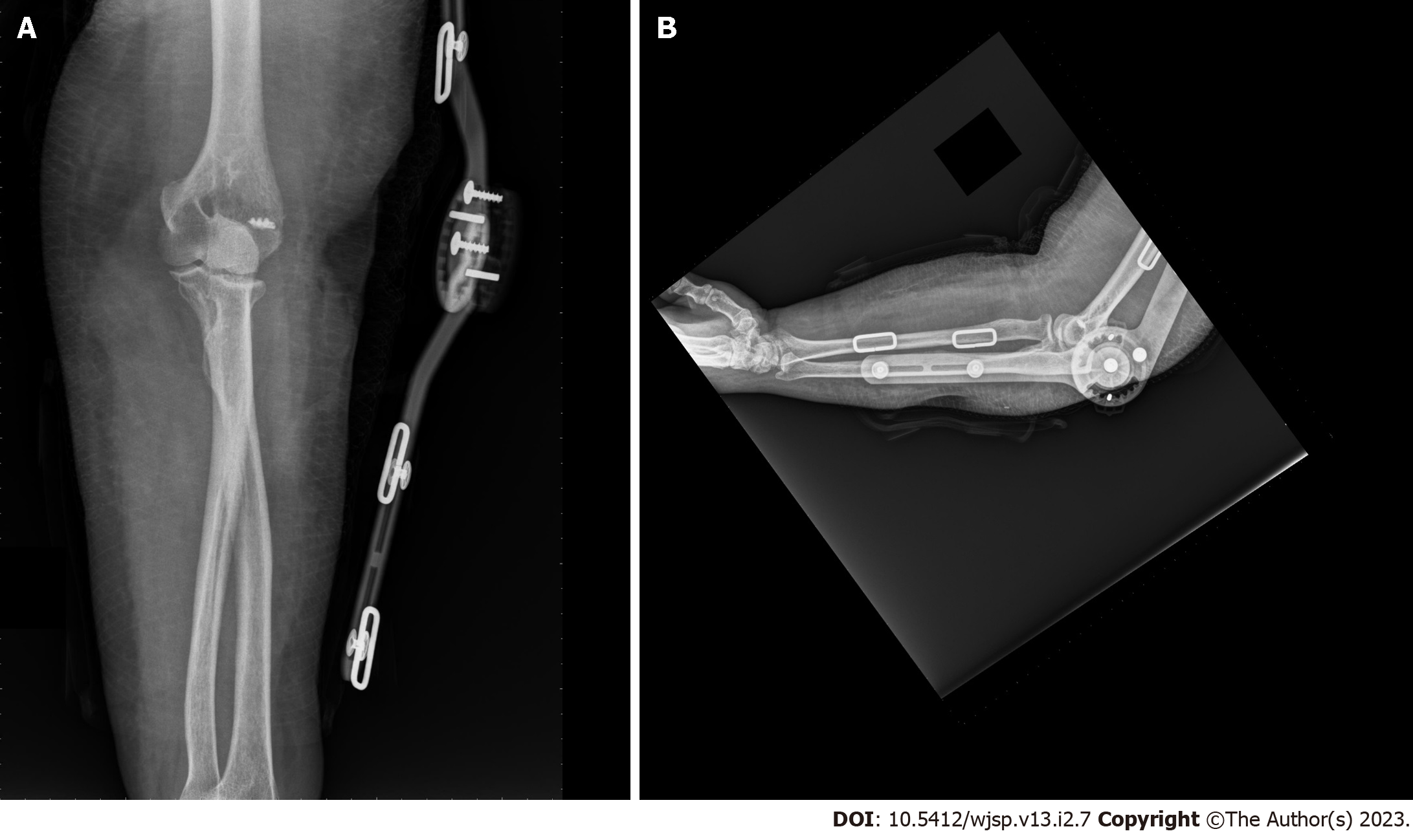
Figure 4 Roentgenographs after surgery.
The elbow is in a brace. A: Anteroposterior; B: Lateral.
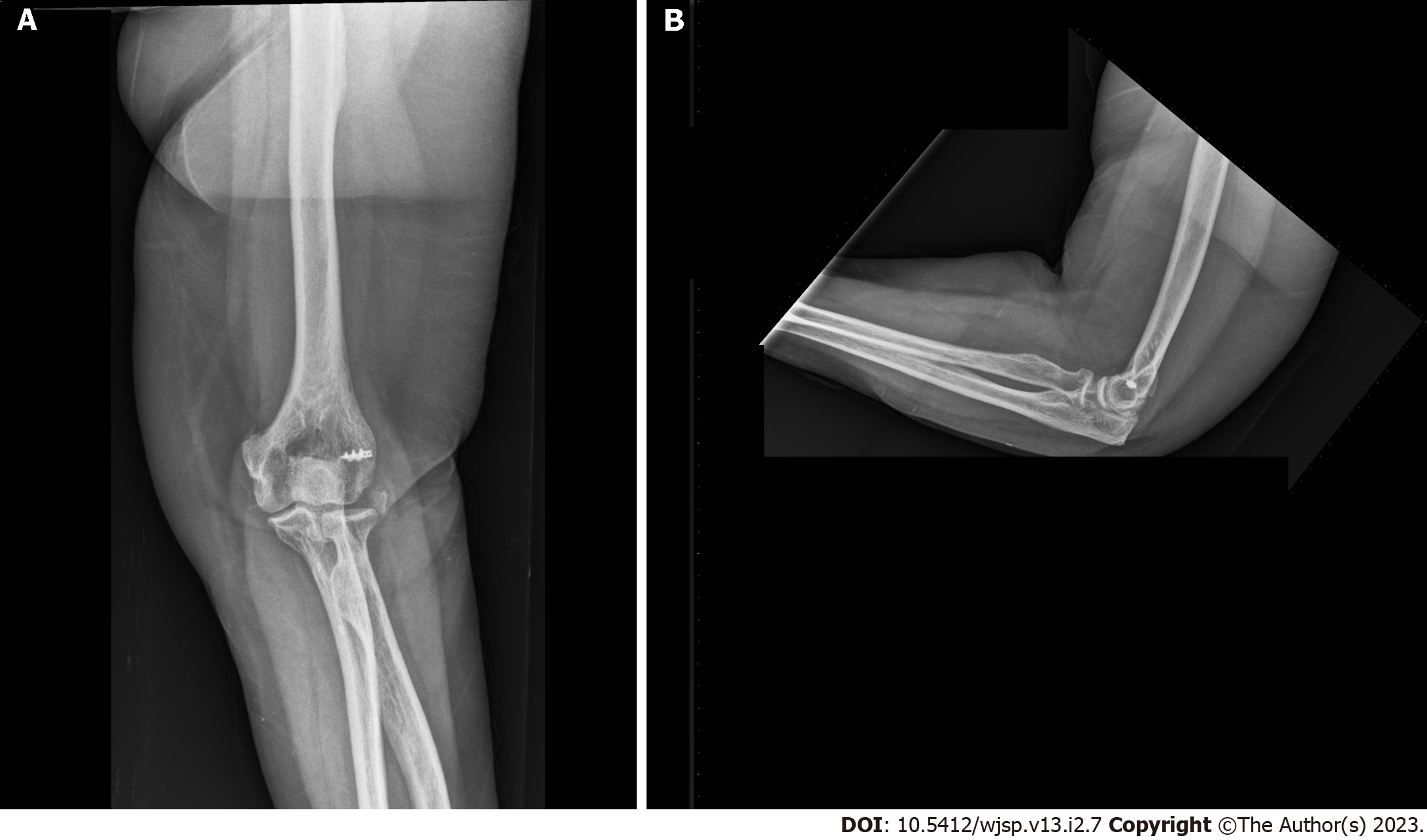
Figure 5 Roentgenographs at the end of two years.
A: Anteroposterior; B: Lateral.
- Citation: Albayrak M. Simple lateral elbow dislocation: A case report. World J Surg Proced 2023; 13(2): 7-13
- URL: https://www.wjgnet.com/2219-2832/full/v13/i2/7.htm
- DOI: https://dx.doi.org/10.5412/wjsp.v13.i2.7


