Copyright
©The Author(s) 2022.
World J Surg Proced. Nov 24, 2022; 12(2): 13-19
Published online Nov 24, 2022. doi: 10.5412/wjsp.v12.i2.13
Published online Nov 24, 2022. doi: 10.5412/wjsp.v12.i2.13
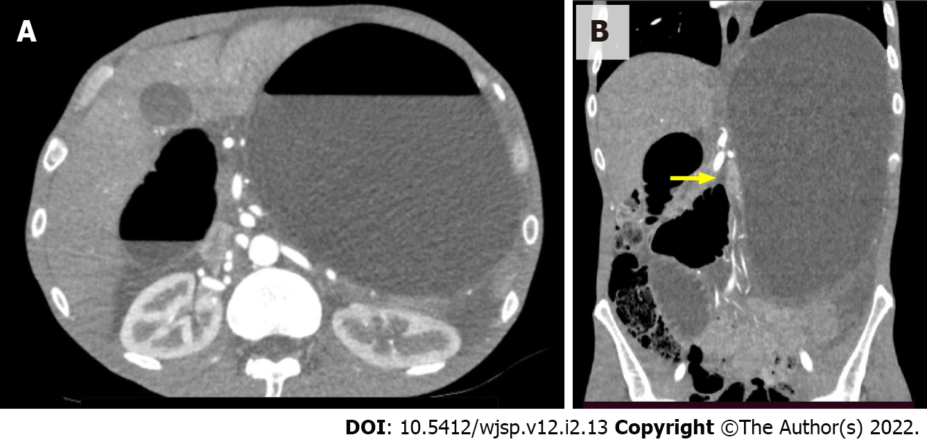
Figure 1 Computed tomography images.
A: Axial computed tomography image showing significant gastric and duodenal distension with formation of air–fluid levels; B: Coronal computed tomography image showing significant gastric and duodenal distension with an abrupt reduction in bowel caliber at the duodenojejunal flexure (yellow arrow).
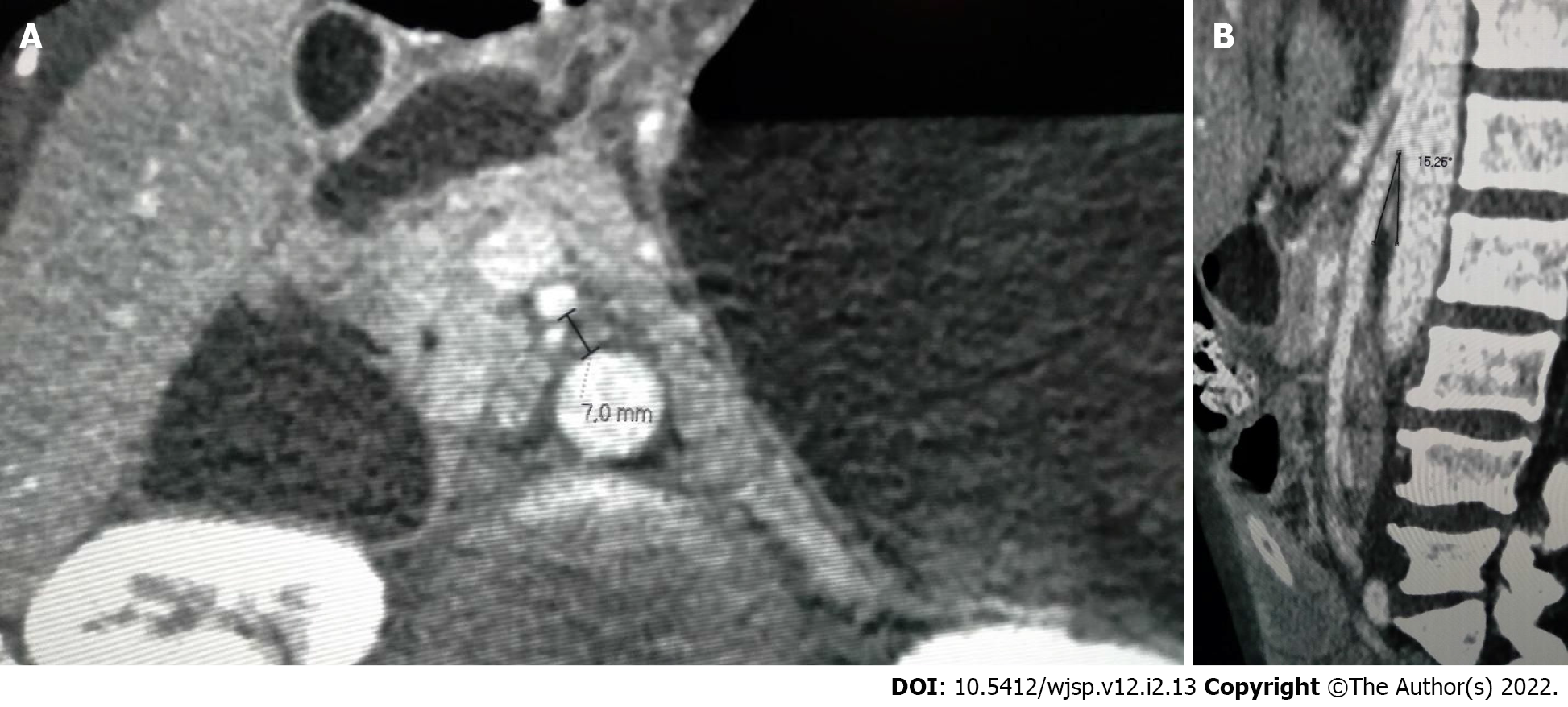
Figure 2 Computed tomography images.
A: Axial computed tomography image showing a distance between the aorta and the superior mesenteric artery of 7 mm (reference > 10 mm); B: Sagittal computed tomography image showing an aortomesenteric angle of 16.26o (reference > 25o).
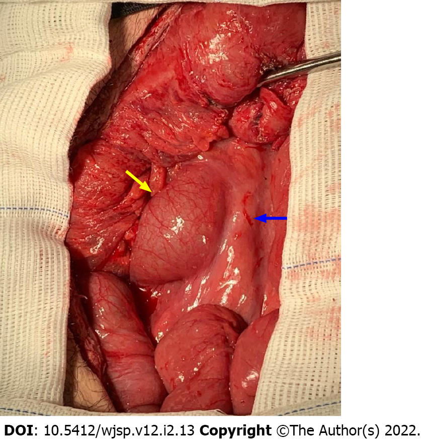
Figure 3
View of intraoperative findings at laparotomy, with marked distention of the duodenum (yellow arrow) up to the point where its third part crosses the superior mesenteric artery (blue arrow), where an abrupt reduction in bowel caliber is seen.
- Citation: Barros LCTR, Santos BMRTD, Ferreira GSA, Gomes VMDS, Vieira LPB. Superior mesenteric artery syndrome in a patient with esophageal stenosis: A case report. World J Surg Proced 2022; 12(2): 13-19
- URL: https://www.wjgnet.com/2219-2832/full/v12/i2/13.htm
- DOI: https://dx.doi.org/10.5412/wjsp.v12.i2.13


