Copyright
©The Author(s) 2021.
World J Surg Proced. Dec 30, 2021; 11(1): 1-9
Published online Dec 30, 2021. doi: 10.5412/wjsp.v11.i1.1
Published online Dec 30, 2021. doi: 10.5412/wjsp.v11.i1.1
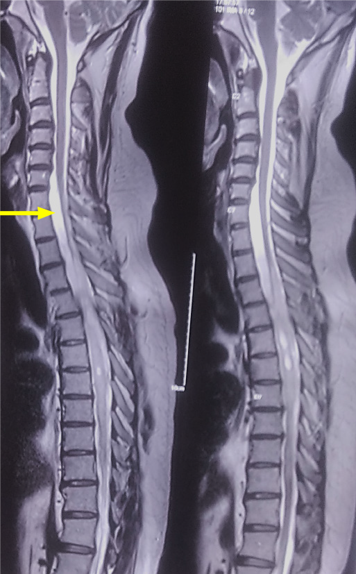
Figure 1 T2 sagittal magnetic resonance imaging sequence showing a multiloculated syrinx from D3-D11.
Arrow points to a subtle loculation of cerebrospinal fluid anterior to the lower cervical and upper dorsal cord.
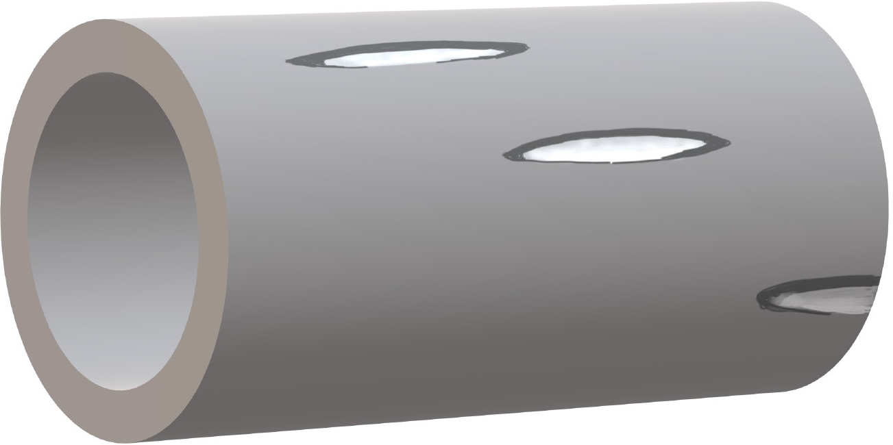
Figure 2
Shunt conduits are prepared and ends fenestrated in a helical fashion to ensure adequate flow of cerebrospinal fluid and minimize the risk of obstruction while also maintaining structural stability.
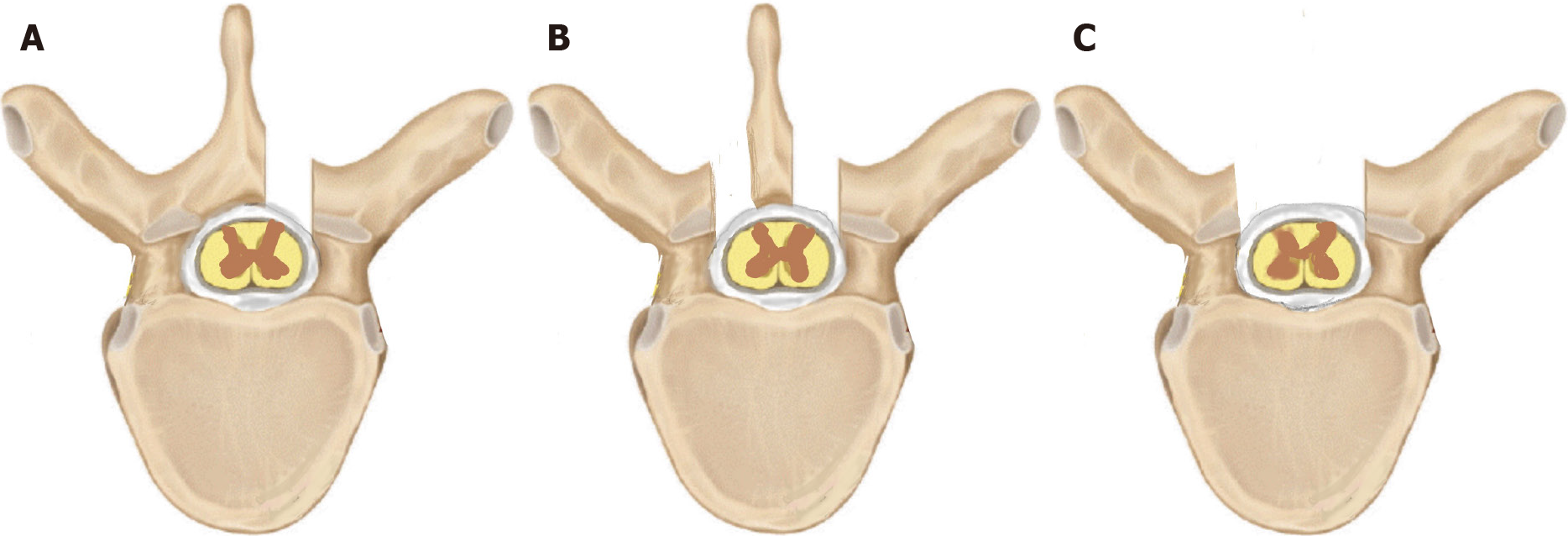
Figure 3 Various approaches to the thecal sac.
A unilateral fenestration (A) may suffice for unilateral theco-thecal shunts whereas a bilateral fenestration (B) or complete laminectomy (C) may be employed in bilaterally placed conduits.
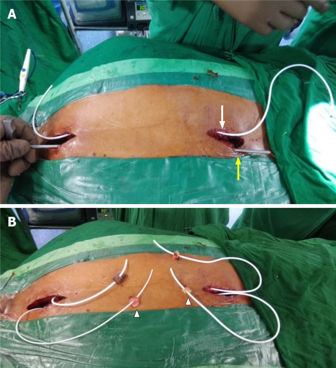
Figure 4 Connecting passages created by subcutaneous tunneling with the help of an adult ventriculoperitoneal shunt tunneller.
A: One after another, both conduits were positioned in the subcutaneous plane on either side of the midline, using an adult ventriculoperitoneal shunt tunneller. The right conduit is seen in place (white arrow) and the tunneller is in use to position the left shunt (yellow arrow); B: Both conduits were placed in the subcutaneous plane and emerged on either side of the incision with fat pads (arrowheads) placed a few centimeters from the tip to help seal the dural entry.
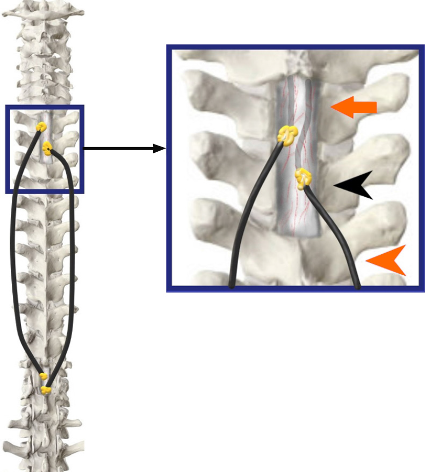
Figure 5 The tubes have been inserted into the theca on both sides of the midline approximately 7-10 mm apart (orange arrowhead).
An adequate intradural length is ensured (orange arrow) and fat grafts are lowered (black arrowhead) over a fibrin seal to augment the sealing (Inset Image).
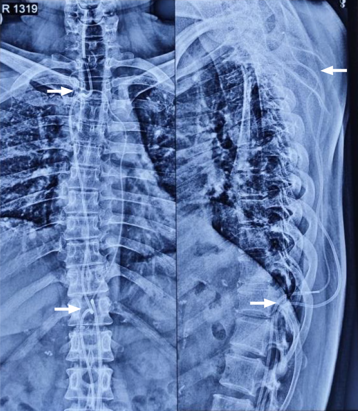
Figure 6
Postoperative plain spine radiographs (AP and lateral) confirming the intraspinal position above at D1-D2 level and below at D11 level (white arrows) along with the continuous subcutaneous track of the conduits and intrathecal portion visible.
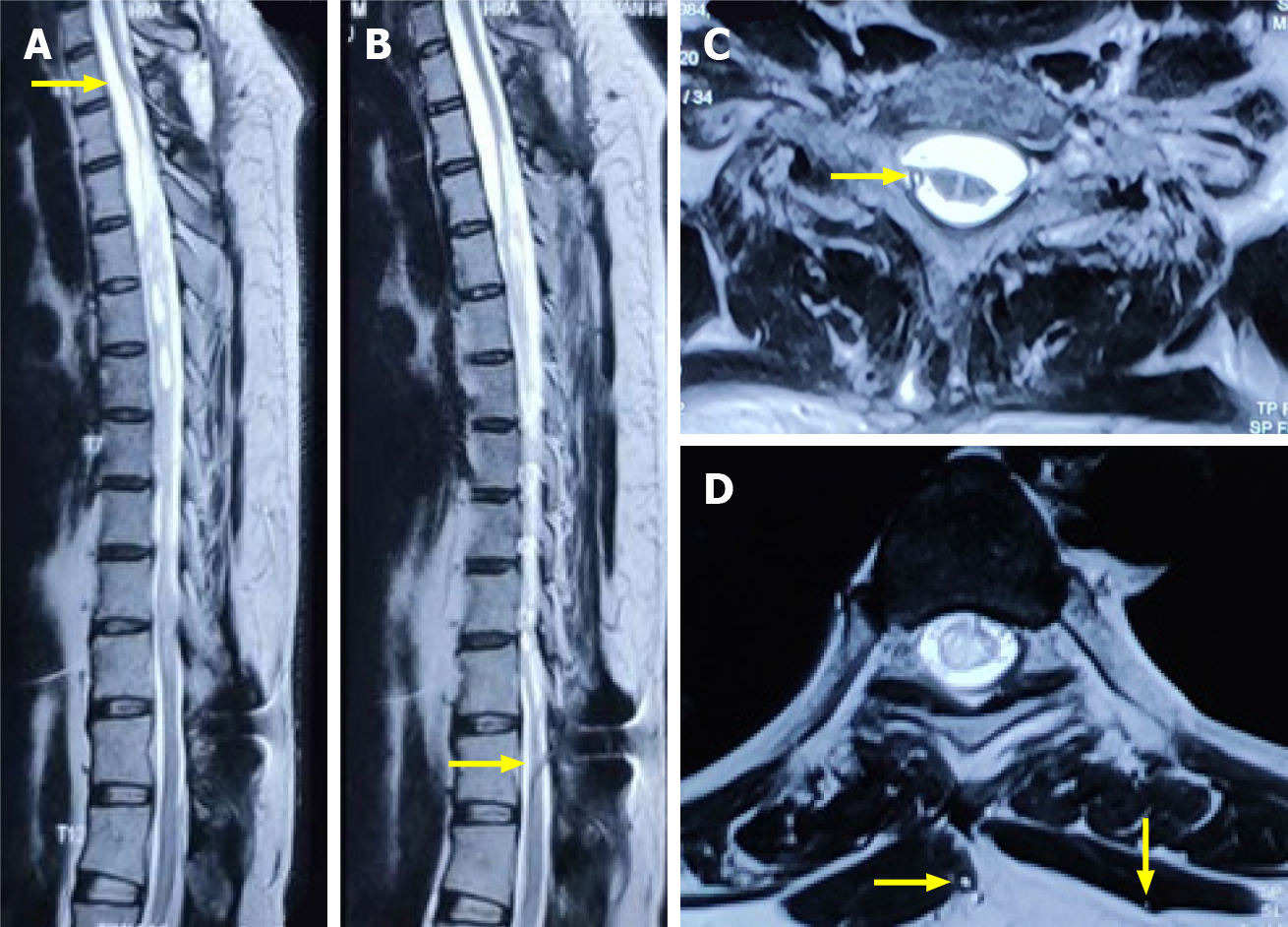
Figure 7 Magnetic resonance imaging images.
T2 weighted sagittal (A and B) and axial magnetic resonance imaging sequences (C and D) at a 4-mo interval showed the upper entry of conduit into the thecal space above and below the level of the syrinx (yellow arrows). No interval change was noted in the size of the syrinx, although a significant clinical and functional improvement was noted.
- Citation: Bhatjiwale M, Bhatjiwale M. Theco-thecal bypass technique elucidating a novel procedure and perspective on treatment of post-arachnoiditis syringomyelia: A case report. World J Surg Proced 2021; 11(1): 1-9
- URL: https://www.wjgnet.com/2219-2832/full/v11/i1/1.htm
- DOI: https://dx.doi.org/10.5412/wjsp.v11.i1.1


