Copyright
©The Author(s) 2015.
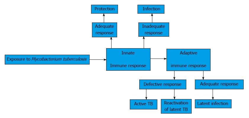
Figure 1 Innate and adaptive immune responses in the Mycobacterium tuberculosis infection.
TB: Tuberculosis.
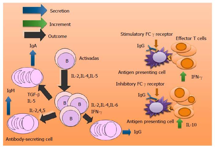
Figure 2 Cytokines involved in antibody secretion in the Mycobacterium tuberculosis infection.
Interaction of IgG with stimulatory or inhibitor receptors and the secretion of cytokines. TGF-β: Transforming growth factor-beta; IL: Interleukin; IFN-γ:Interferon-γ.
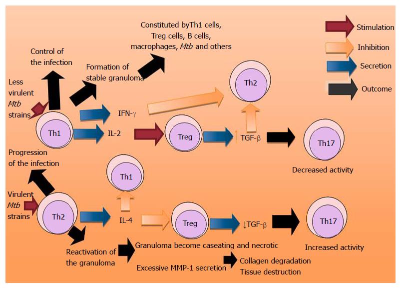
Figure 3 Participation of Th1, Th2, Th17 and T reg phenotypes in the evolution of the Mycobacterium tuberculosis infection.
TGF-β: Transforming growth factor-beta; IL: Interleukin; IFN-γ: Interferon-γ.
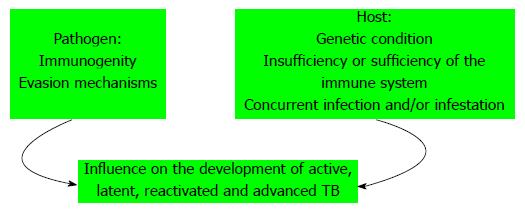
Figure 4 Factors involved in the evolution of Mycobacterium tuberculosis infection.
TB: Tuberculosis.
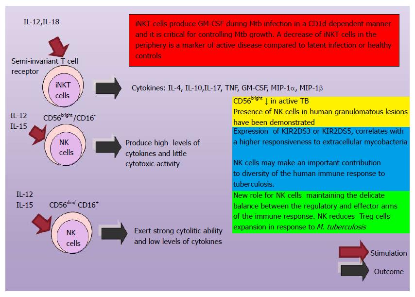
Figure 5 Participation of invariant natural killer T and natural killer cells in the control of Mycobacterium tuberculosis infection.
iNKT: Invariant natural killer T; CD: Clusters of differentiation; MIP: Macrophage inflammatory protein; GM-CSF: Granulocyte macrophage colony-stimulating factor; TB: Tuberculosis; NK: Natural killer; IL: Interleukin.
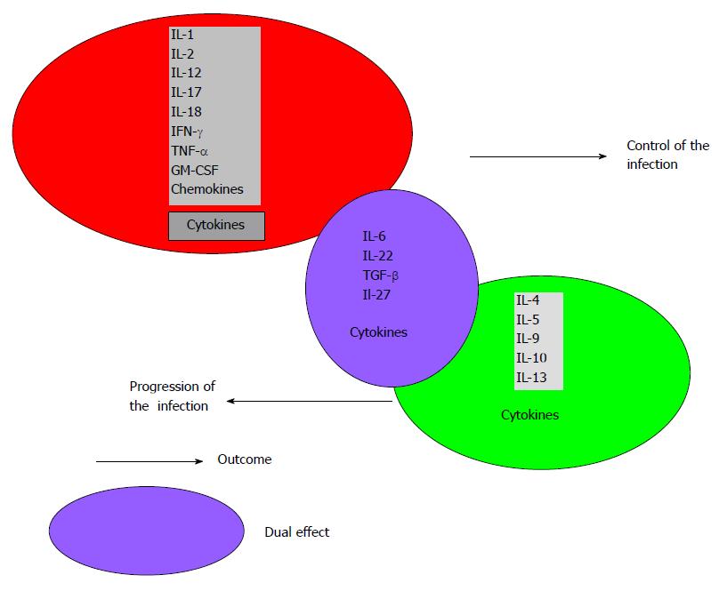
Figure 6 Cytokines in the control and progression of the Mycobacterium tuberculosis infection.
The concentrations of cytokines, microenvironment and other factors contribute to modify the outcome. TGF-β: Transforming growth factor-beta; GM-CSF: Granulocyte macrophage colony-stimulating factor; IL: Interleukin; IFN-γ:Interferon-γ; TNF-α; Tumor necrosis factor α.
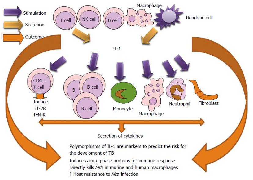
Figure 7 Biological effects of interleukin 1 in the Mycobacterium tuberculosis infection.
TB: Tuberculosis; NK: Natural killer; IL: Interleukin; CD: Clusters of differentiation; Mtb: Mycobacterium tuberculosis; IFN: Interferon.
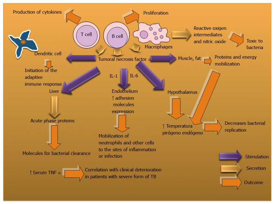
Figure 8 Biological actions of Tumor necrosis factor α to control Mycobacterium tuberculosis infection.
TB: Tuberculosis; IL: Interleukin.
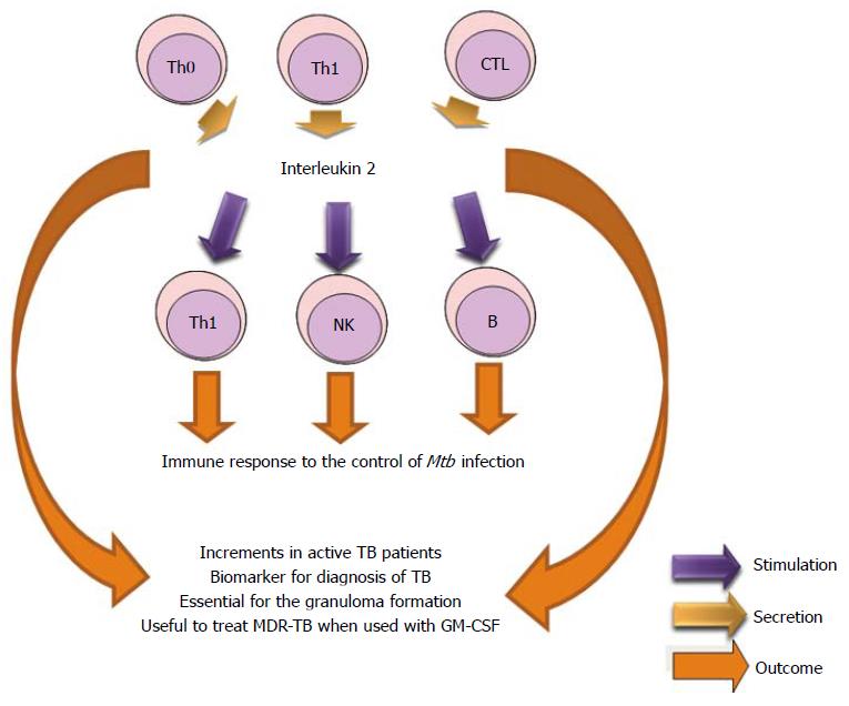
Figure 9 Role of IL-2 in Mycobacterium tuberculosis infection.
TB: Tuberculosis; MDR-TB: Multidrug resistant tuberculosis; GM-CSF: Granulocyte macrophage colony-stimulating factor; NK: Natural killer; MDR-TB: Multi drug resistant tuberculosis.
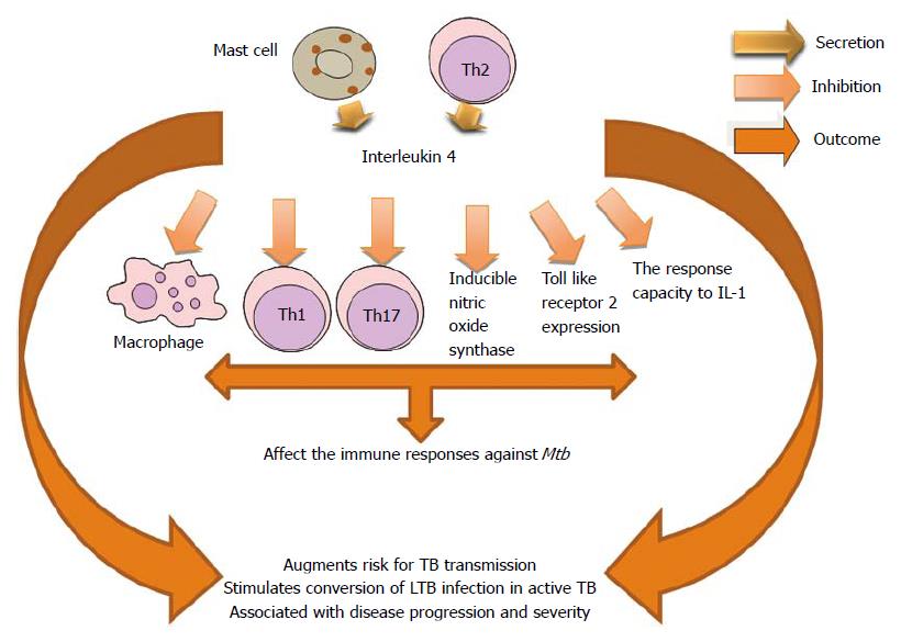
Figure 10 Participation of interleukin-4 in Mycobacterium tuberculosis infection.
TB: Tuberculosis; LTB: Latent tuberculosis.
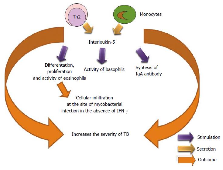
Figure 11 Participation of interleukin-5 in the Mycobacterium tuberculosis infection.
TB: Tuberculosis; IFN-γ: Interferon-γ.
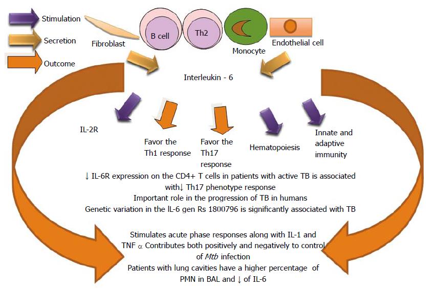
Figure 12 Role of interleukin-6 in the Mycobacterium tuberculosis infection.
TB: Tuberculosis; PMN: Polymorphonuclear neutrophils; IL: Interleukin.
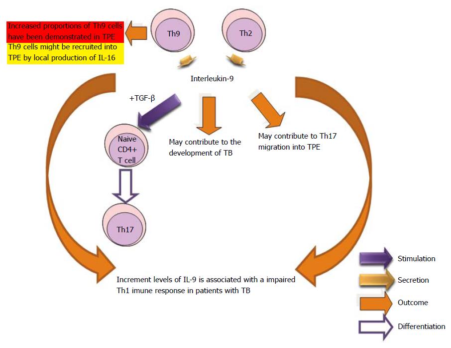
Figure 13 Participation of interleukin-9 in the Mycobacterium tuberculosis infection.
TB: Tuberculosis; TPE: Tuberculous pleural effusion; TGF-β: Transforming growth factor-beta; CD: Clusters of differentiation; IL: Interleukin.
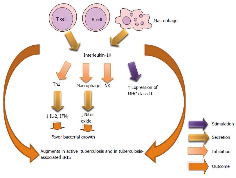
Figure 14 Influence of interleukin-10 in the control of Mycobacterium tuberculosis infection.
IRIS: Immune reconstitution inflammatory syndrome; IFNγ:Interferon γ; NK: Natural killer; IL: Interleukin.
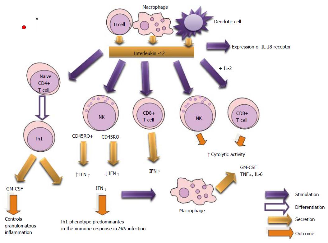
Figure 15 Biological actions of interleukin-12 to control of the Mycobacterium tuberculosis infection.
GM-CSF: Granulocyte macrophage colony-stimulating factor; NK: Natural killer; CD: Clusters of differentiation; IL: Interleukin; IFN γ:Interferon γ.
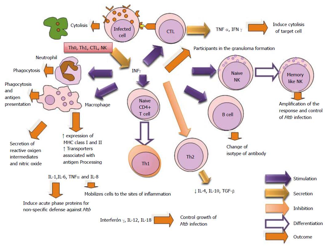
Figure 16 Biological actions of Interferon-γ in Mycobacterium tuberculosis infection.
TGF-β: Transforming growth factor-beta; NK: Natural killer; CTL: Cytotoxic t lymphocyte; CD: Clusters of differentiation; IL: Interleukin; CTL: Computation tree logic; Th1: T helper type 1.
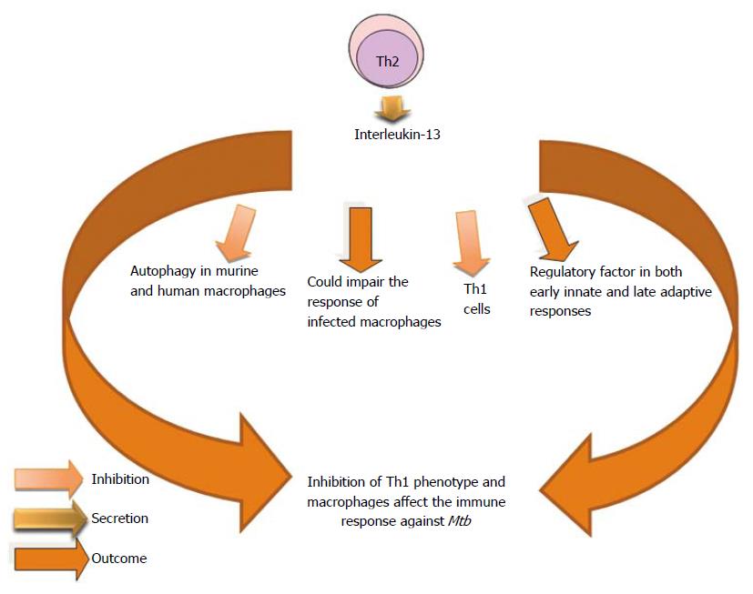
Figure 17 Participation of interleukin-13 in Mycobacterium tuberculosis infection.
Th1: T helper type 1; Th2: T helper type 2; Mtb: Mycobacterium tuberculosis.
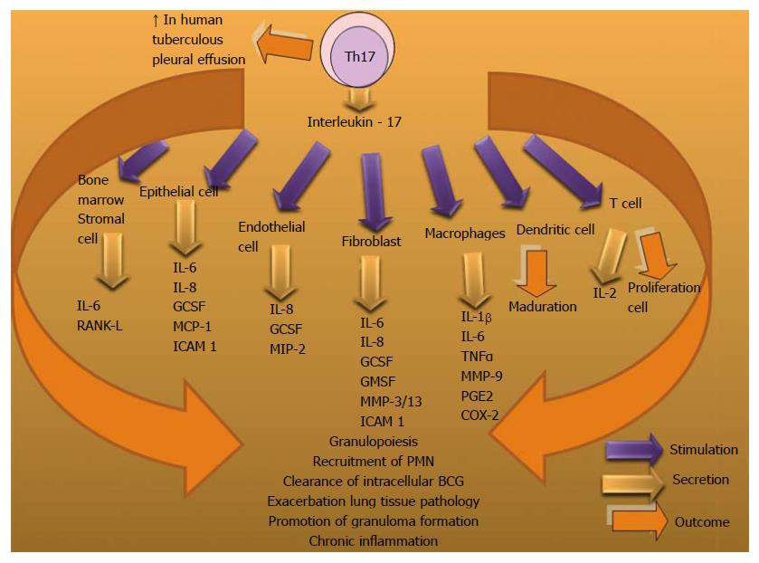
Figure 18 Interleukin-17 in the control of Mycobacterium tuberculosis infection.
MCP: Monocyte chemoattractant protein; RANK-L: Receptor activator of NF-Κb ligand; MMP-9: Metalloproteinases-9; GCSF: Granulocyte colony stimulating factor; MIP-2: Macrophage inflammatory protein-2; BCG: Bacillus calmette guérin; PGE2: Prostaglandin e-2; COX-2: Cyclooxigenase-2; ICAM 1: Intercellular adhesion molecules-1; IL: Interleukin.
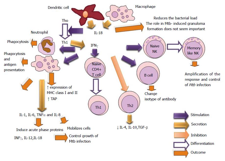
Figure 19 Role of interleukin-18 in Mycobacterium tuberculosis infection.
TGF-β: Transforming growth factor-beta; NK: Natural killer; TAP: Transporters associated with antigen processing; CD: Clusters of differentiation; IL: Interleukin.
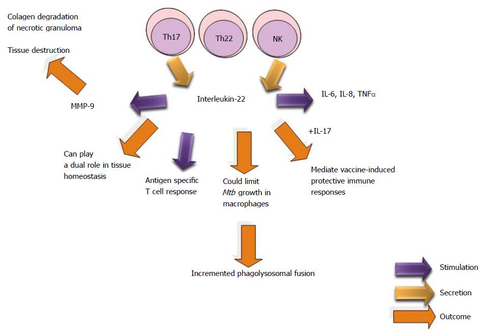
Figure 20 Biological Effects of interleukin-22 in the Mycobacterium tuberculosis infection.
MMP-9: Metalloproteinases-9; NK: Natural killer; IL: Interleukin.
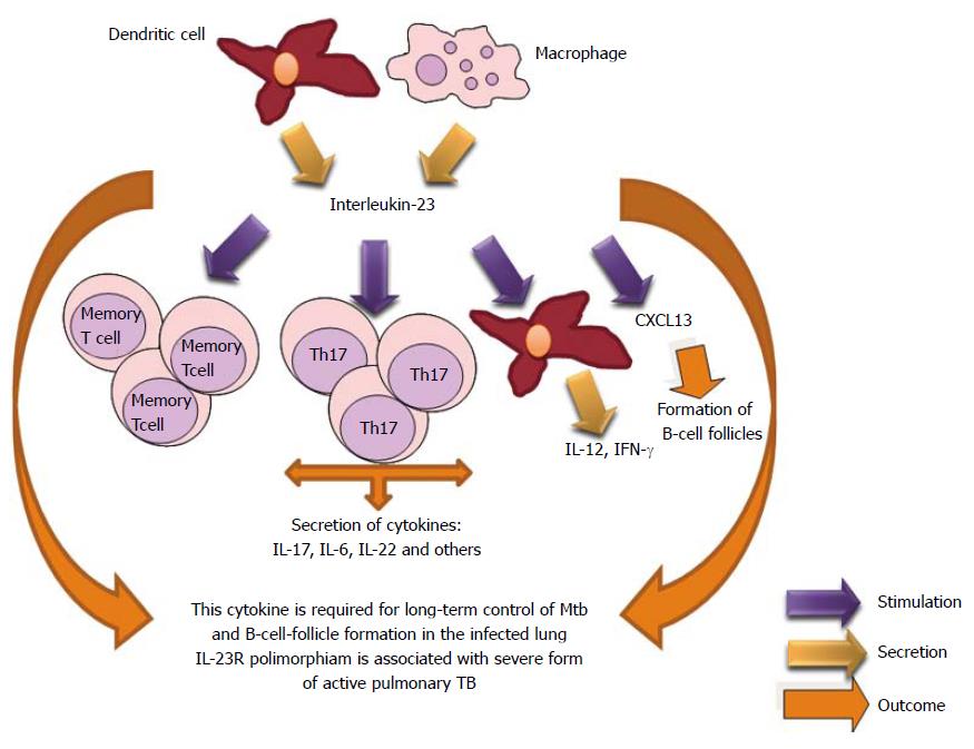
Figure 21 Participation of interleukin-23 in Mycobacterium tuberculosis infection.
TB: Tuberculosis; IL: Interleukin; IFN-γ: Interferon-γ.
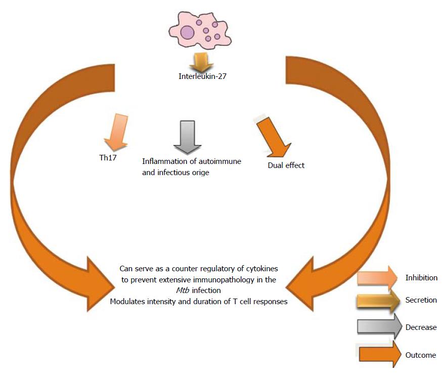
Figure 22 Role of interleukin-27 in Mycobacterium tuberculosis infection.
Th17: T helper type 17; Mtb: Mycobacterium tuberculosis.
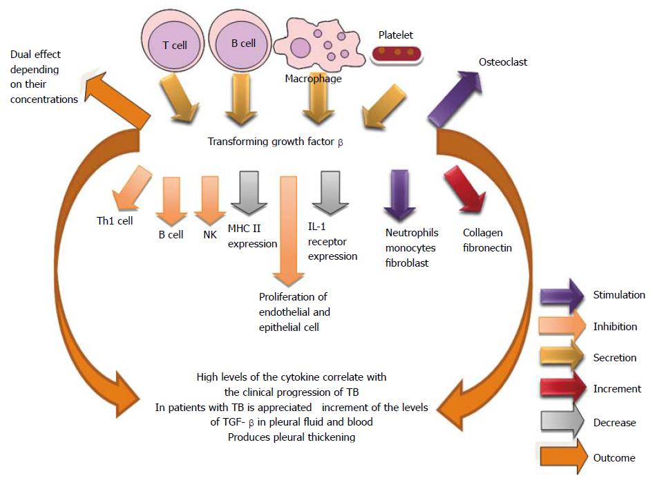
Figure 23 Transforming growth factor-beta in the control of Mycobacterium tuberculosis infection.
TB: Tuberculosis; Th1: T helper type 1; TGF-β: Transforming growth factor-beta; NK: Natural killer; IL: Interleukin.
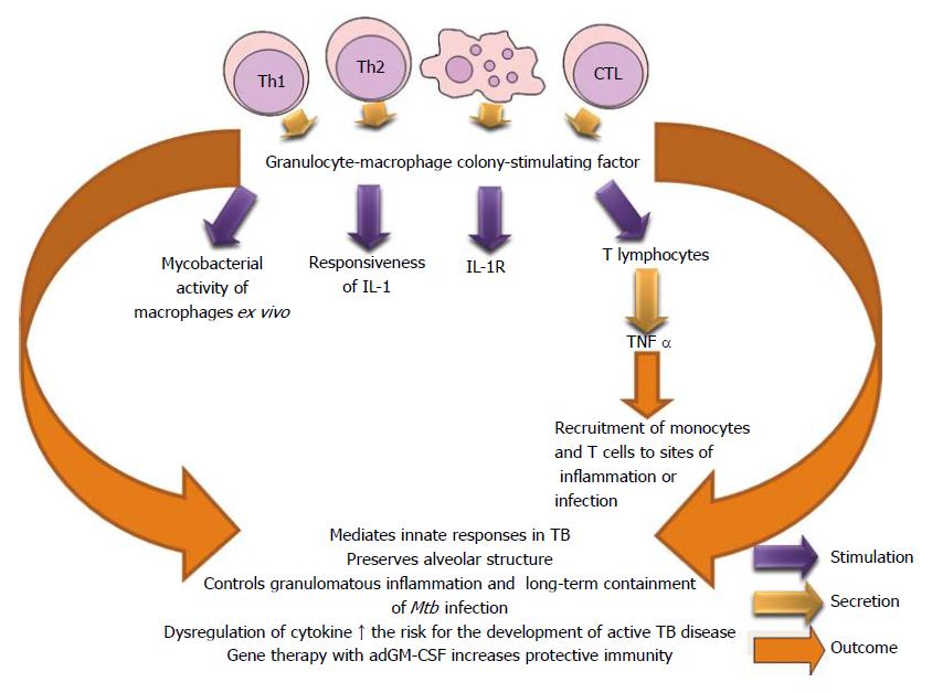
Figure 24 Influence of granulocyte macrophage colony-stimulating factor in the control of Mycobacterium tuberculosis infection.
GM-CSF: Granulocyte macrophage colony-stimulating factor; IL: Interleukin; Mtb: Mycobacterium tuberculosis; TB: Tuberculosis; TNF: Tumor necrosis factor.
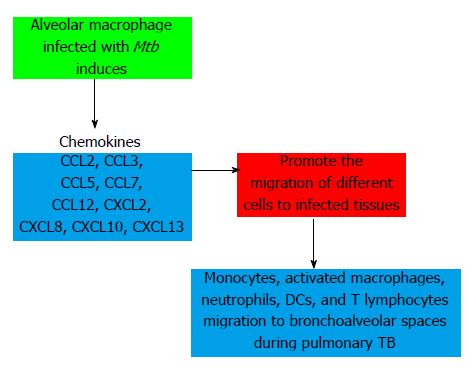
Figure 25 Participation of chemokines in pulmonary tuberculosis.
TB: Tuberculosis; DCs: Dendritic cells.
-
Citation: Romero-Adrian TB, Leal-Montiel J, Fernández G, Valecillo A. Role of cytokines and other factors involved in the
Mycobacterium tuberculosis infection. World J Immunol 2015; 5(1): 16-50 - URL: https://www.wjgnet.com/2219-2824/full/v5/i1/16.htm
- DOI: https://dx.doi.org/10.5411/wji.v5.i1.16


