Copyright
©The Author(s) 2017.
World J Clin Urol. Mar 24, 2017; 6(1): 30-33
Published online Mar 24, 2017. doi: 10.5410/wjcu.v6.i1.30
Published online Mar 24, 2017. doi: 10.5410/wjcu.v6.i1.30
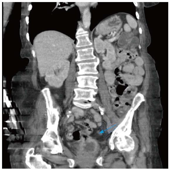
Figure 1 Computed tomography abdomen/pelvis demonstrating the enterovesical fistula from dome of bladder to terminal ileum (arrow).
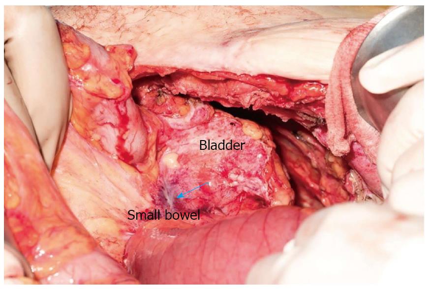
Figure 2 An enterovesical fistula is demonstrated (arrow) intraoperatively.
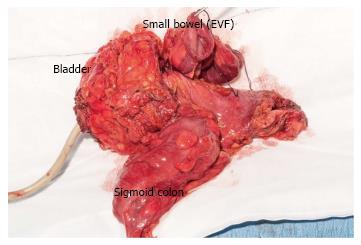
Figure 3 En-bloc resection including radical cystectomy, small bowel resection and sigmoid colectomy.
EVF: Enterovesical fistulae.
- Citation: Ng ZQ, Low WKW, Jr S, Subramanian P, Stein J. Radical cystectomy and en-bloc resection of enterovesical fistula from bladder cancer. World J Clin Urol 2017; 6(1): 30-33
- URL: https://www.wjgnet.com/2219-2816/full/v6/i1/30.htm
- DOI: https://dx.doi.org/10.5410/wjcu.v6.i1.30


