Copyright
©The Author(s) 2017.
Figure 1 Filling defect on retropyelogram, showing typical “goblet sign” due to R ureteral urothelial cell carcinoma.
Figure 2 Central mass of upper tract urothelial cell carcinoma on left kidney, seen as renal sinus mass.
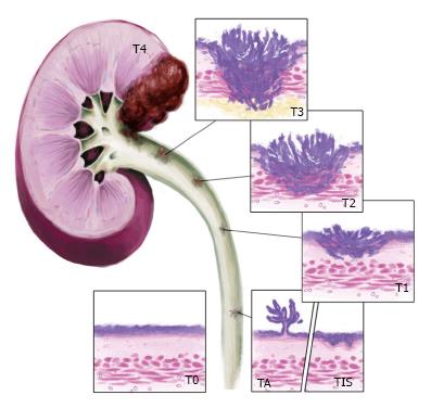
Figure 3 Pathologic stage of upper tract urothelial cell carcinoma.
Courtesy of third year medical student at West Virginia University, Mike Tran.
Figure 4 Obtaining tumor specimen for pathologic stage.
Figure 5 Tumor resection using ureteral resectoscope with loop.
- Citation: Choi K, McCafferty R, Deem S. Contemporary management of upper tract urothelial cell carcinoma. World J Clin Urol 2017; 6(1): 1-9
- URL: https://www.wjgnet.com/2219-2816/full/v6/i1/1.htm
- DOI: https://dx.doi.org/10.5410/wjcu.v6.i1.1


