Copyright
©The Author(s) 2015.
World J Clin Urol. Nov 24, 2016; 5(3): 93-96
Published online Nov 24, 2016. doi: 10.5410/wjcu.v5.i3.93
Published online Nov 24, 2016. doi: 10.5410/wjcu.v5.i3.93
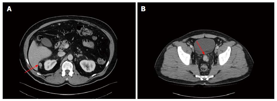
Figure 1 Axial computed tomography image of the abdomen.
A: Red arrow pointing to soft tissue mass in between liver and right kidney; B: Red arrow pointing to soft tissue mass between bladder and rectum.
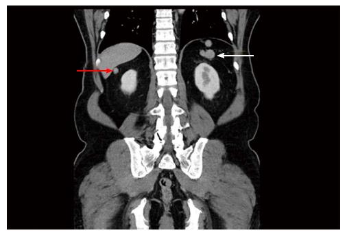
Figure 2 Coronal computed tomography image of the pelvis.
White arrow pointing to nodular soft tissue mass in left upper quadrant, where discrete spleen was not visualised; red arrow pointing to soft tissue mass beneath liver.
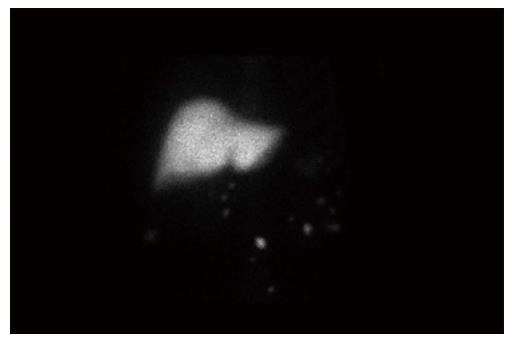
Figure 3 Nuclear scintigraphy image of this patient demonstrating uptake in the ectopic tissue seen in the pelvis and beneath the liver on the computed tomography images, confirming they are ectopic splenic tissue.
Liver shows normal tracer uptake.
- Citation: Foreman D, Plagakis SA. Splenunculi mimicking metastases in a patient with locally advanced prostate cancer. World J Clin Urol 2016; 5(3): 93-96
- URL: https://www.wjgnet.com/2219-2816/full/v5/i3/93.htm
- DOI: https://dx.doi.org/10.5410/wjcu.v5.i3.93


