Copyright
©The Author(s) 2016.
World J Clin Urol. Mar 24, 2016; 5(1): 37-44
Published online Mar 24, 2016. doi: 10.5410/wjcu.v5.i1.37
Published online Mar 24, 2016. doi: 10.5410/wjcu.v5.i1.37
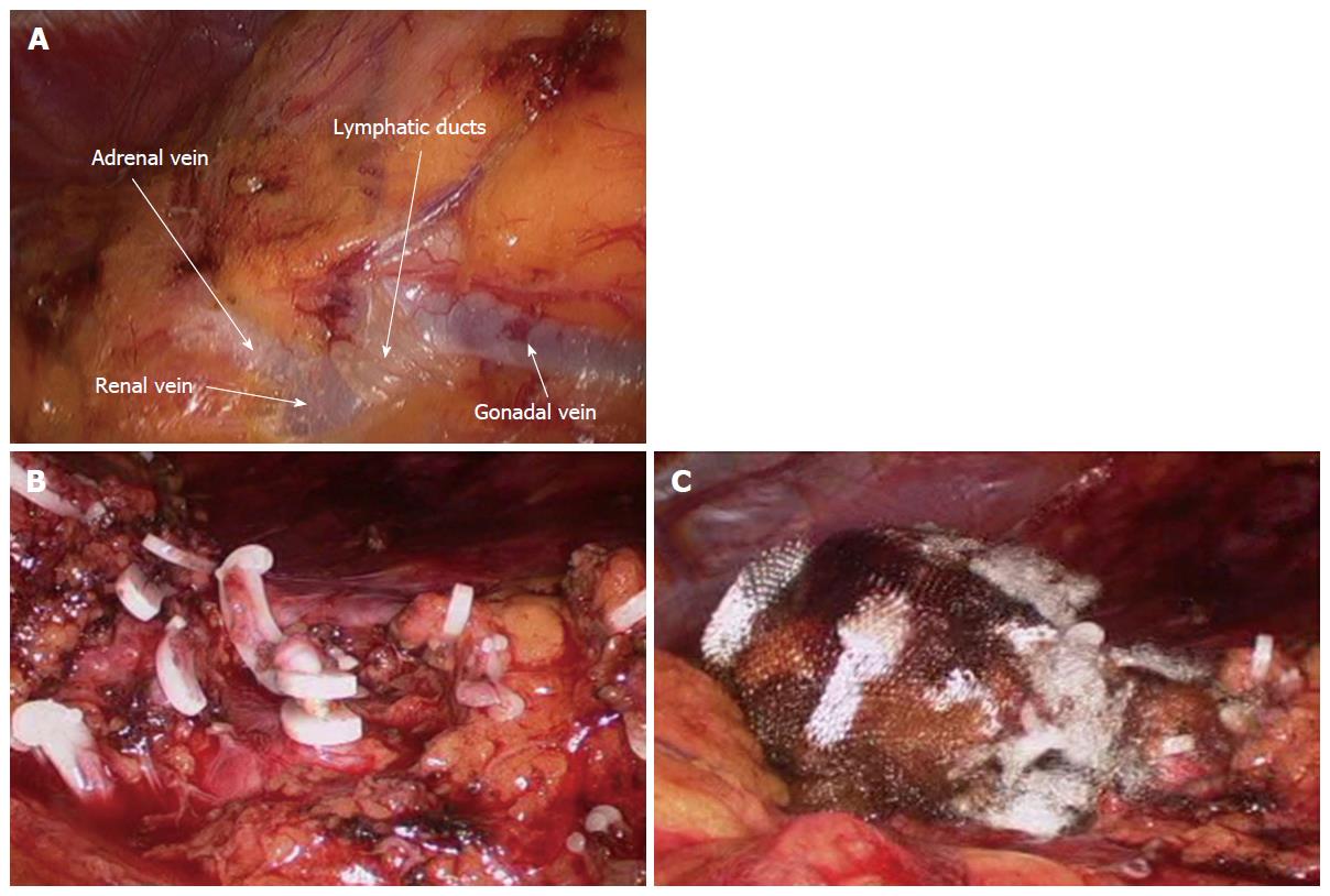
Figure 1 Intraoperative image of the hilar area during left-sided laparoscopic donor nephrectomy.
A: Prominent lymphatic ducts cross the renal vein; B: The perihilar and retroperitoneal fatty tissue is meticulously clipped; C: The hilar area is completely sealed using surgicel and fibrin glue.
- Citation: Kim BS, Kwon TG. Chylous ascites in laparoscopic renal surgery: Where do we stand? World J Clin Urol 2016; 5(1): 37-44
- URL: https://www.wjgnet.com/2219-2816/full/v5/i1/37.htm
- DOI: https://dx.doi.org/10.5410/wjcu.v5.i1.37


