Copyright
©The Author(s) 2016.
World J Clin Urol. Mar 24, 2016; 5(1): 1-10
Published online Mar 24, 2016. doi: 10.5410/wjcu.v5.i1.1
Published online Mar 24, 2016. doi: 10.5410/wjcu.v5.i1.1
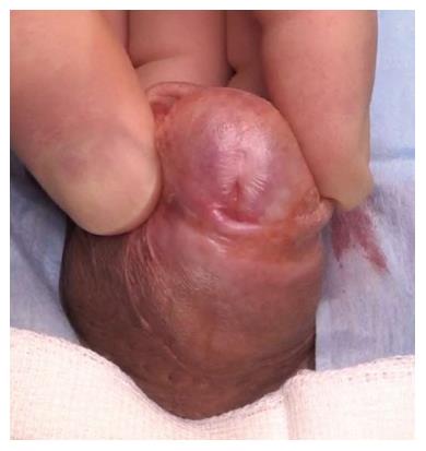
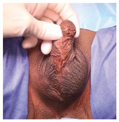
Figure 1 Typical appearance of lichen sclerosus with scarring on the glans, loss of the normal contour of the glans and coronal sulcus, meatal regression and stenosis.
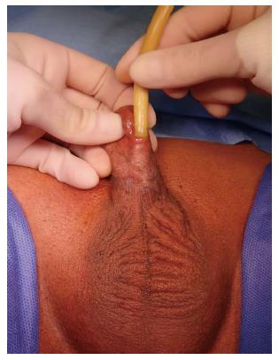
Figure 2 Urethral stricture after failed mid-penile hypospadias reconstruction.
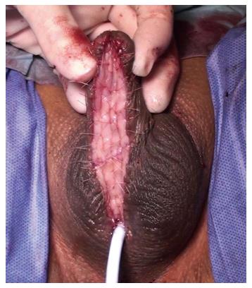
Figure 3 Operative image showing a complete first stage full-length penile urethroplasty using bilateral buccal mucosal grafts quilted dorsally to create a neo-urethral plate of adequate calibre to be retubularised in the second stage.
Figure 4 Operative image showing (A) harvesting of a sublingual graft and (B) bilateral sublingual graft donor sites closed primarily.
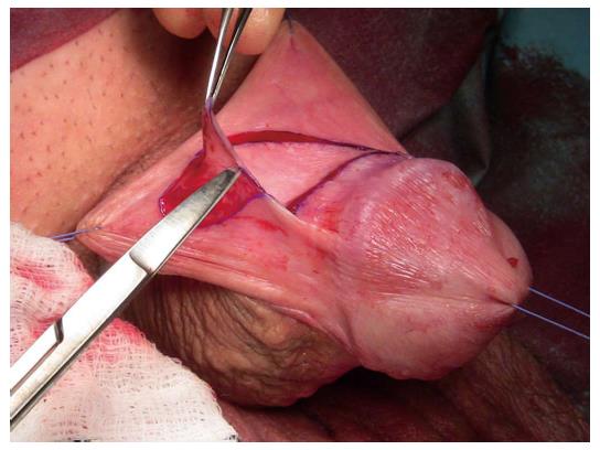
Figure 5 Operative image showing a preputial skin free graft being harvested.
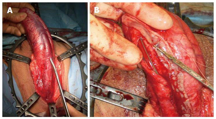
Figure 6 Kulkarni technique for long penile strictures via a transperineal approach with (A) invagination of the penis and (B) dorsolateral placement of the sublingual graft.
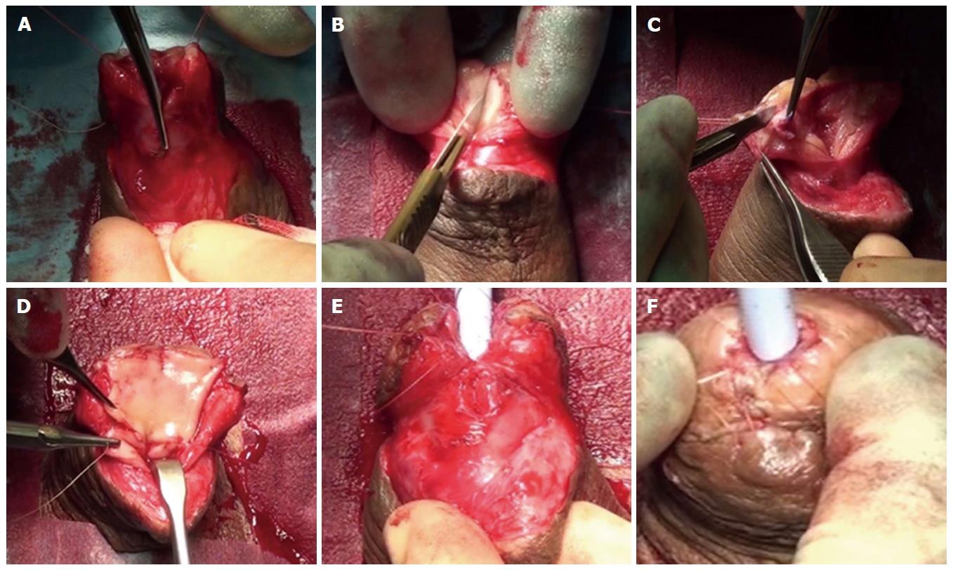
Figure 7 “Two-in-one” stage approach for a distal penile urethral stricture using oral mucosal graft.
A: Ventral stricturotomy; B: Glans cleft deepened; C: Spongiofibrosis excised; D: Neo-urethral plate created using oral mucosa; E and F: Retubularised in layers in one stage.
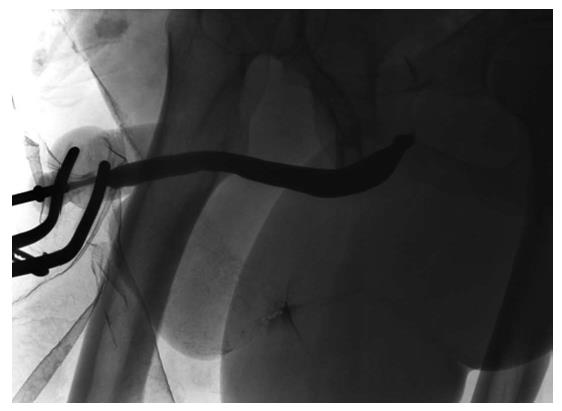
Figure 8 Ascending urethrogram showing a short penile urethral stricture limited to the fossa navicularis.
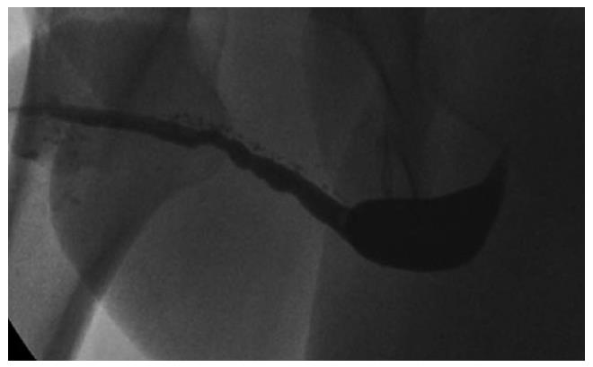
Figure 9 Entire penile and distal bulbar urethra affected by a Lichen Sclerosus-related stricture.
Figure 10 Multiple urethrocutaneous fistulae after failed hypospadias repair.
- Citation: Campos-Juanatey F, Bugeja S, Ivaz SL, Frost A, Andrich DE, Mundy AR. Management of penile urethral strictures: Challenges and future directions. World J Clin Urol 2016; 5(1): 1-10
- URL: https://www.wjgnet.com/2219-2816/full/v5/i1/1.htm
- DOI: https://dx.doi.org/10.5410/wjcu.v5.i1.1


