Copyright
©2014 Baishideng Publishing Group Inc.
World J Clin Urol. Nov 24, 2014; 3(3): 376-379
Published online Nov 24, 2014. doi: 10.5410/wjcu.v3.i3.376
Published online Nov 24, 2014. doi: 10.5410/wjcu.v3.i3.376
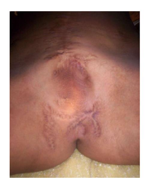
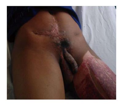
Figure 1 Preoperative view demonstrating perineal urethrostomy.
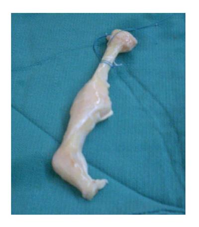
Figure 2 Cadaveric bone allograft construct, using metacarpal and proximal phalanx.
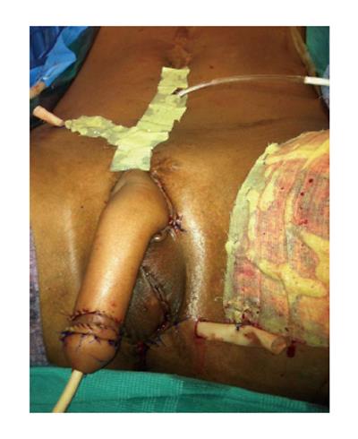
Figure 3 Immediate post-operative view.
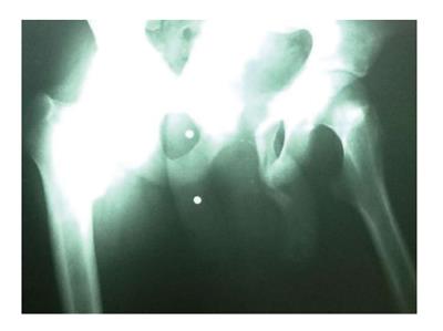
Figure 4 X-ray views demonstrating bone allograft construct within the substance of the neophallus.
Figure 5 Six months follow-up demonstrating viable neophallus.
- Citation: Edens JW, Tran T, Eidelson S, Askari M, Salgado CJ. Pre-fabricated radial forearm phalloplasty with cadaveric bone graft. World J Clin Urol 2014; 3(3): 376-379
- URL: https://www.wjgnet.com/2219-2816/full/v3/i3/376.htm
- DOI: https://dx.doi.org/10.5410/wjcu.v3.i3.376


