Copyright
©2014 Baishideng Publishing Group Inc.
World J Clin Urol. Nov 24, 2014; 3(3): 272-282
Published online Nov 24, 2014. doi: 10.5410/wjcu.v3.i3.272
Published online Nov 24, 2014. doi: 10.5410/wjcu.v3.i3.272
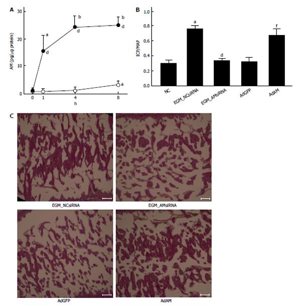
Figure 1 Production of adrenomedullin by adipose tissue-derived stem cells, and effects of knockdown and overexpression of adrenomedullin on the function and histology of the penis.
A: Adipose tissue-derived stem cells produce adrenomedullin especially when they were cultured in a medium containing growth factors for vascular endothelial cells (VECs); adipose tissue-derived stem cells (ADSCs) were cultured in endothelial basal medium (EBM: white circles) or endothelial growth medium (EGM: black circles) that contains growth factors for VECs. Medium was replaced with serum-free medium and incubated for the indicated periods. Adrenomedullin (AM) accumulated in the medium was measured. aP < 0.05 vs 0 h, bP < 0.01 vs 0 h and dP < 0.01 vs EBM culture at each time point (n = 6 per group); B: Effect of knockdown and overexpression of AM on erectile function. ADSCs were infected with lentivirus expressing negative control siRNA (LV_NCsiRNA) that is predicted not to target any known vertebrate gene or lentivirus expressing AM siRNA (LV_AMsiRNA). ADSCs were cultured in EGM for 1 wk, and those LV_NCsiRNA-infected ADSCs (EGM_NCsiRNA) and LV_AMsiRNA-infected ADSCs (EGM_AMsiRNA) were injected in the cavernous body of STZ-induced diabetic rats. ICP was measured 4 wk after the ADSCs injection. An adenovirus expressing AM (AdAM) or adenovirus expressing green fluorescent protein (AdGFP) was also injected into the cavernous body of STZ-induced diabetic rats, and ICP was measured 4 wk after the infection. Nontreated STZ-injected diabetic rats were used as the negative control (NC). Bar graphs show ICP/MAP (n = 5 per group). aP < 0.001 vs NC, dP < 0.001 vs EGM_NCsiRNA injection and fP < 0.001 vs AdGFP infection; C: Effect of knockdown and overexpression of AM on the morphology of the cavernous body. Experiments were performed in the same way as described in the legend for Figure 1B. The cavernous body was stained by the Elastica van Gieson method. The histology of the root portion of the penis (longitudinal section) is shown. Bars are 200 μm. Note that the size of trabeculae of the cavernous body is smaller when EGM_AMsiRNA or AdGFP was injected into the penis of diabetic rats, compared with when EGM_NCsiRNA or AdAM was injected into the penis. ICP: Intracavernous pressure; STZ: Streptozotocin; MAP: Mean arterial pressure.
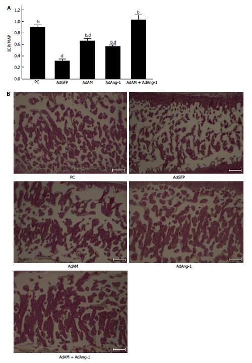
Figure 2 Effects of adrenomedullin and angiopoietin-1 on the function and morphology of the penis.
A: Effects of adenovirus expressing adrenomedullin (AdAM), adenovirus expressing angiopoietin-1 (AdAng-1) and AdAM plus AdAng-1 infection on erectile function. These adenoviruses were infected into the cavernous body of STZ-induced diabetic rats. Adenovirus expressing green fluorescent protein (AdGFP) was also infected as the negative control. Age-matched Wistar rats were used as the positive control (PC). Bar graphs demonstrate ICP/MAP (n = 6 per group). bP < 0.001 vs AdGFP infection and dP < 0.001 vs PC; B: Histological analysis of the cavernous body after adenoviral infection. Elastica van Gieson staining of the cavernous body isolated from age-matched Wistar rats (PC), and STZ-induced diabetic rats infected with AdGFP, AdAM, AdAng-1, and AdAM plus AdAng-1. The histology of the root portion of the penis (longitudinal section) is shown. Bars are 300 μm. Note that the size of trabeculae of the cavernous body is small when AdGFP is injected into the penis of diabetic rats, and that the size is restored to a similar level as observed in the age-matched control group when AdAM and/or AdAng-1 are injected. ICP: Intracavernous pressure; STZ: Streptozotocin; MAP: Mean arterial pressure.
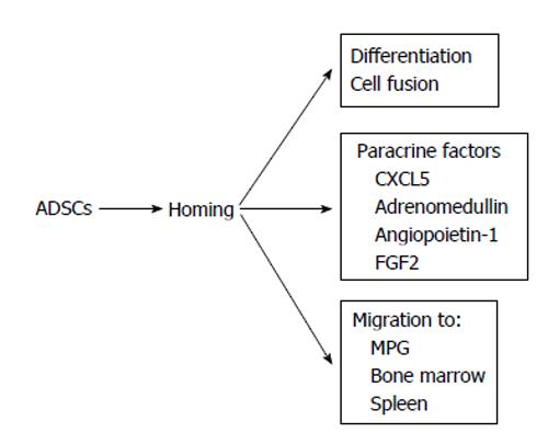
Figure 3 Possible mechanisms by which adipose tissue-derived stem cells stimulate the recovery of erectile function.
FGF2: Fibroblast growth factor 2; MPG: Major pelvic ganglia; ADSCs: Adipose tissue-derived stem cells; CXCL5: Chemokine (C-X-C motif) ligand 5.
- Citation: Suzuki E, Nishimatsu H, Homma Y. Stem cell therapy for erectile dysfunction. World J Clin Urol 2014; 3(3): 272-282
- URL: https://www.wjgnet.com/2219-2816/full/v3/i3/272.htm
- DOI: https://dx.doi.org/10.5410/wjcu.v3.i3.272


