Copyright
©2014 Baishideng Publishing Group Inc.
World J Clin Urol. Nov 24, 2014; 3(3): 218-226
Published online Nov 24, 2014. doi: 10.5410/wjcu.v3.i3.218
Published online Nov 24, 2014. doi: 10.5410/wjcu.v3.i3.218
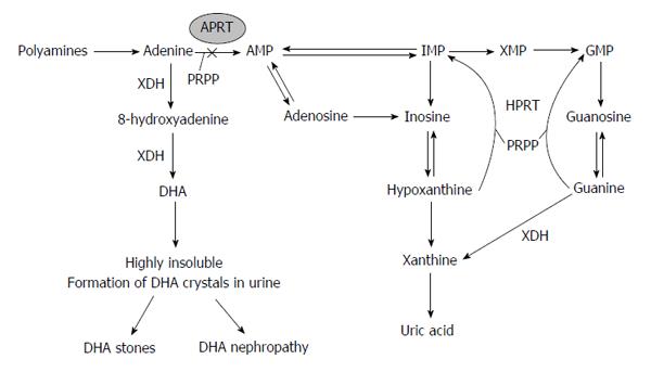
Figure 1 Biochemical pathway of purine metabolism and mechanisms of adenine phosphoribosyltransferase deficiency.
DHA: 2,8-dihydroxyadenine; APRT: Adenine phosphoribosyltransferase; HPRT: Hypoxanthine phosphoribosyltransferase; PRPP: Phosphoribosyl-pyrophosphate; IMP: Inosine monophosphate; XDH: Xanthine dehydrogenase enzyme; AMP: Adenosine monophosphate; GMP: Guanosine monophosphate; XMP: Xanthosine monophosphate.
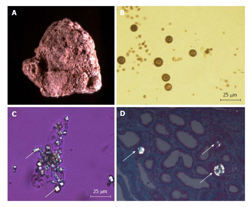
Figure 2 Stones and crystals of 2,8-dihydroxyadenine.
A: Typical aspect of a 2,8-dihydroxyadenine (DHA) stone, showing a rough and humpy surface and a reddish-brown color that turns grey when drying. The stone is friable and sections show porosities and beige to brown color; B: Light microscopy aspect of DHA crystals in urine (non-polarized image), showing a round shape and a reddish-brown color; C: Urine DHA crystals in polarized light, showing typical central Maltese cross pattern (arrows); D: Periodic acid-Schiff of kidney biopsy under polarized light in a patient with adenine phosphoribosyltransferase deficiency, showing crystals within the renal tubules (arrows) (× 400). Fourier transform infrared microscopy study of the biopsy demonstrated that crystals were composed of DHA.
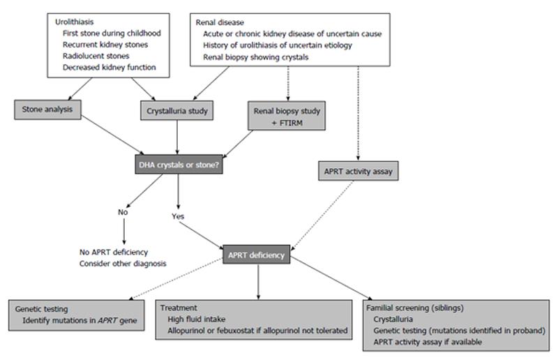
Figure 3 Algorithm for diagnosis and management of adenine phosphoribosyltransferase deficiency.
This clinical algorithm summarizes situations where adenine phosphoribosyltransferase (APRT) deficiency should be suspected, testing strategies and management of the disease. FTIRM: Fourier transform infrared microscopy; DHA: 2,8-dihydroxyadenine.
- Citation: Bollée G, Daudon M, Ceballos-Picot I. Adenine phosphoribosyltransferase deficiency: Leave no stone unturned. World J Clin Urol 2014; 3(3): 218-226
- URL: https://www.wjgnet.com/2219-2816/full/v3/i3/218.htm
- DOI: https://dx.doi.org/10.5410/wjcu.v3.i3.218


