Copyright
©2014 Baishideng Publishing Group Inc.
World J Clin Urol. Jul 24, 2014; 3(2): 66-80
Published online Jul 24, 2014. doi: 10.5410/wjcu.v3.i2.66
Published online Jul 24, 2014. doi: 10.5410/wjcu.v3.i2.66
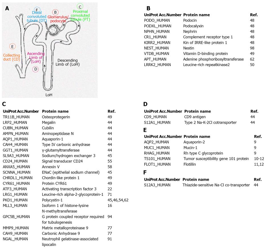
Figure 1 Schematics of Nephron showing the urinary extracellular vesicle proteins identified in the different segments.
A: Core; B: Glomerulus/podocyte; C: Proximal convoluted tubule (PT); D: Ascending Limb of (LoH); E: Collecting duct (CD); F: Distal convoluted tubule (DT). UniProt Acc.Number: UniProt Database Accession Number. LoH: Loop of Henle.
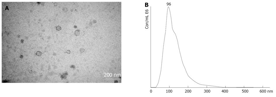
Figure 2 (A) Transmission electron microscopy and (B) nanoparticle tracking analysis images of urinary extracellular vesicles isolated by the sucrose cushion ultracentrifugation method.
- Citation: Pocsfalvi G, Stanly C, Vilasi A, Fiume I, Tatè R, Capasso G. Employing extracellular vesicles for non-invasive renal monitoring: A captivating prospect. World J Clin Urol 2014; 3(2): 66-80
- URL: https://www.wjgnet.com/2219-2816/full/v3/i2/66.htm
- DOI: https://dx.doi.org/10.5410/wjcu.v3.i2.66


