Copyright
©The Author(s) 2015.
World J Clin Pediatr. Nov 8, 2015; 4(4): 66-80
Published online Nov 8, 2015. doi: 10.5409/wjcp.v4.i4.66
Published online Nov 8, 2015. doi: 10.5409/wjcp.v4.i4.66
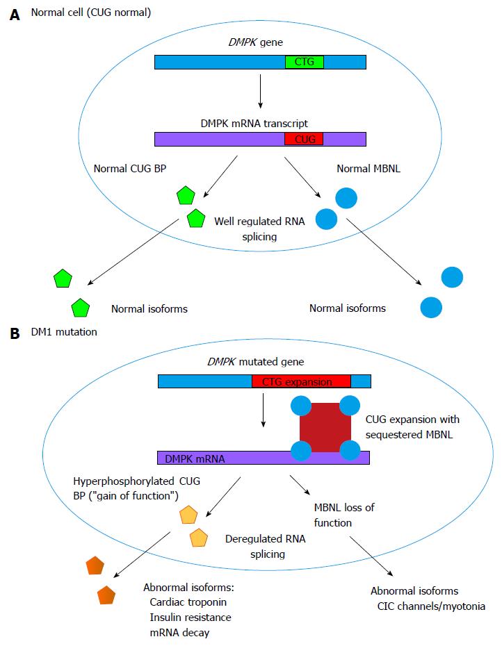
Figure 3 Pathogenic mechanisms in myotonic dystrophy type 1: (A) Normal RNA processing in cell with normal CTG repeats at the myotonic dystrophy type 1 locus; (B) Effects of expanded CTG repeat at the DM1 locus.
A: Normal actions of MBNL and CUG BP in regulating alternative splicing within a cell; B: Pathogenic mechanisms involving MBNL and CUG BP, resulting in deregulated alternative splicing. While DM1 mutation is an untranslated CTG expansion, it is expressed at the RNA level and the CUG binds with two RNA binding proteins (CUGBP and MBNL) to disrupt RNA processing and splicing of other genes. For example, altered splicing of chloride channel and insulin receptor transcripts leads to myotonia and insulin resistance, respectively. DM1: Myotonic dystrophy type 1; MBNL: Musclebind-like protein; CUG BP: CUG binding protein.
- Citation: Ho G, Cardamone M, Farrar M. Congenital and childhood myotonic dystrophy: Current aspects of disease and future directions. World J Clin Pediatr 2015; 4(4): 66-80
- URL: https://www.wjgnet.com/2219-2808/full/v4/i4/66.htm
- DOI: https://dx.doi.org/10.5409/wjcp.v4.i4.66


