Copyright
©The Author(s) 2016.
World J Clin Pediatr. May 8, 2016; 5(2): 228-233
Published online May 8, 2016. doi: 10.5409/wjcp.v5.i2.228
Published online May 8, 2016. doi: 10.5409/wjcp.v5.i2.228
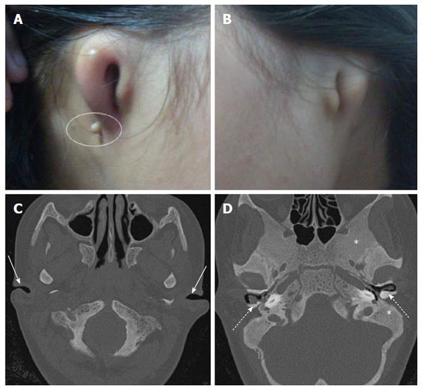
Figure 1 Bilateral dysplastic pinnae.
Note preauricular skin tag on right side (circle). Axial HRCT (C and D) showing bilateral mild narrowing of cartilaginous EAC (arrows) and normal bony EAC (dotted arrow) with ear wax. Also seen are the thickened sclerotic calvarial bones with loss of normal medullary cavity (asterisk). EAC: External auditory canal.
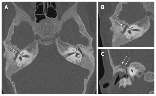
Figure 2 Axial high resolution computed tomography images.
(A and B) showing soft tissue opacification in bilateral middle ears (A) with erosion of head of head of malleus (arrows). Coronal reformatted CT image (C) showing thinning and erosion of tegmen tympani (dotted arrows) secondary to cholesteatoma.
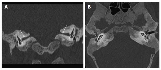
Figure 3 Coronal reformatted high resolution computed tomography.
(A) image showing thickened sclerotic bones causing narrowing of bilateral internal auditory canal (arrows); normal cochlea and vestibule seen in both ears on axial high resolution computed tomography image (B).
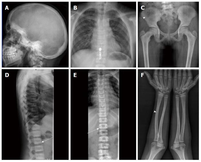
Figure 4 Skeletal survey.
Note generalised increased bone density in all the radiographs (A to F). Calvarial bones as well as skull base are thickened (A), lung fields are normal with sclerotic ribs (B). No paraspinal masses to suggest extra medullary hematopoiesis. Sclerosis of pelvic bones with subtle blurring of cortico-medullary is differentiation in long bones (arrow heads). Sclerosis along vertebral endplates (dotted arrow) leading to “rugger jersy spine” (D and E).
- Citation: Verma R, Jana M, Bhalla AS, Kumar A, Kumar R. Diagnosis of osteopetrosis in bilateral congenital aural atresia: Turning point in treatment strategy. World J Clin Pediatr 2016; 5(2): 228-233
- URL: https://www.wjgnet.com/2219-2808/full/v5/i2/228.htm
- DOI: https://dx.doi.org/10.5409/wjcp.v5.i2.228


