Copyright
©The Author(s) 2015.
World J Clin Pediatr. Nov 8, 2015; 4(4): 120-125
Published online Nov 8, 2015. doi: 10.5409/wjcp.v4.i4.120
Published online Nov 8, 2015. doi: 10.5409/wjcp.v4.i4.120
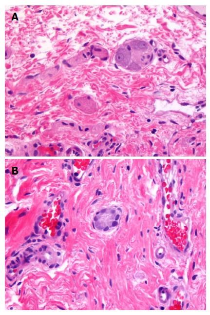
Figure 1 Mature ganglion cells (A) and immature ganglion cells (B).
A: Mature ganglion cells; B: Immature ganglion cells (Hematoxylin and Eosin staining, magnification: 400 × for both photographs).
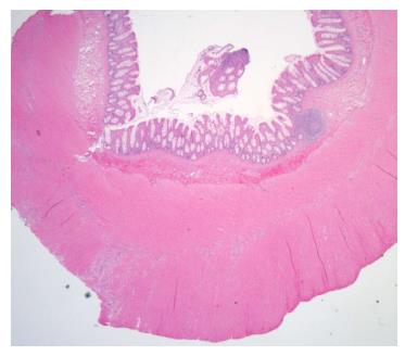
Figure 2 Hirschsprung's disease showing hypertrophy of nerve fibers.
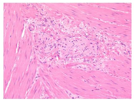
Figure 3 Hirschsprung's disease showing hypertrophy of nerve fibers and lack of ganglion cells.
- Citation: Sergi C. Hirschsprung’s disease: Historical notes and pathological diagnosis on the occasion of the 100th anniversary of Dr. Harald Hirschsprung’s death. World J Clin Pediatr 2015; 4(4): 120-125
- URL: https://www.wjgnet.com/2219-2808/full/v4/i4/120.htm
- DOI: https://dx.doi.org/10.5409/wjcp.v4.i4.120


