Copyright
©The Author(s) 2024.
World J Clin Pediatr. Jun 9, 2024; 13(2): 93341
Published online Jun 9, 2024. doi: 10.5409/wjcp.v13.i2.93341
Published online Jun 9, 2024. doi: 10.5409/wjcp.v13.i2.93341
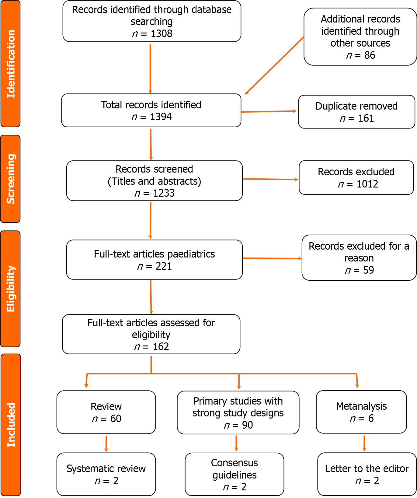
Figure 1
The flow chart of the included studies.
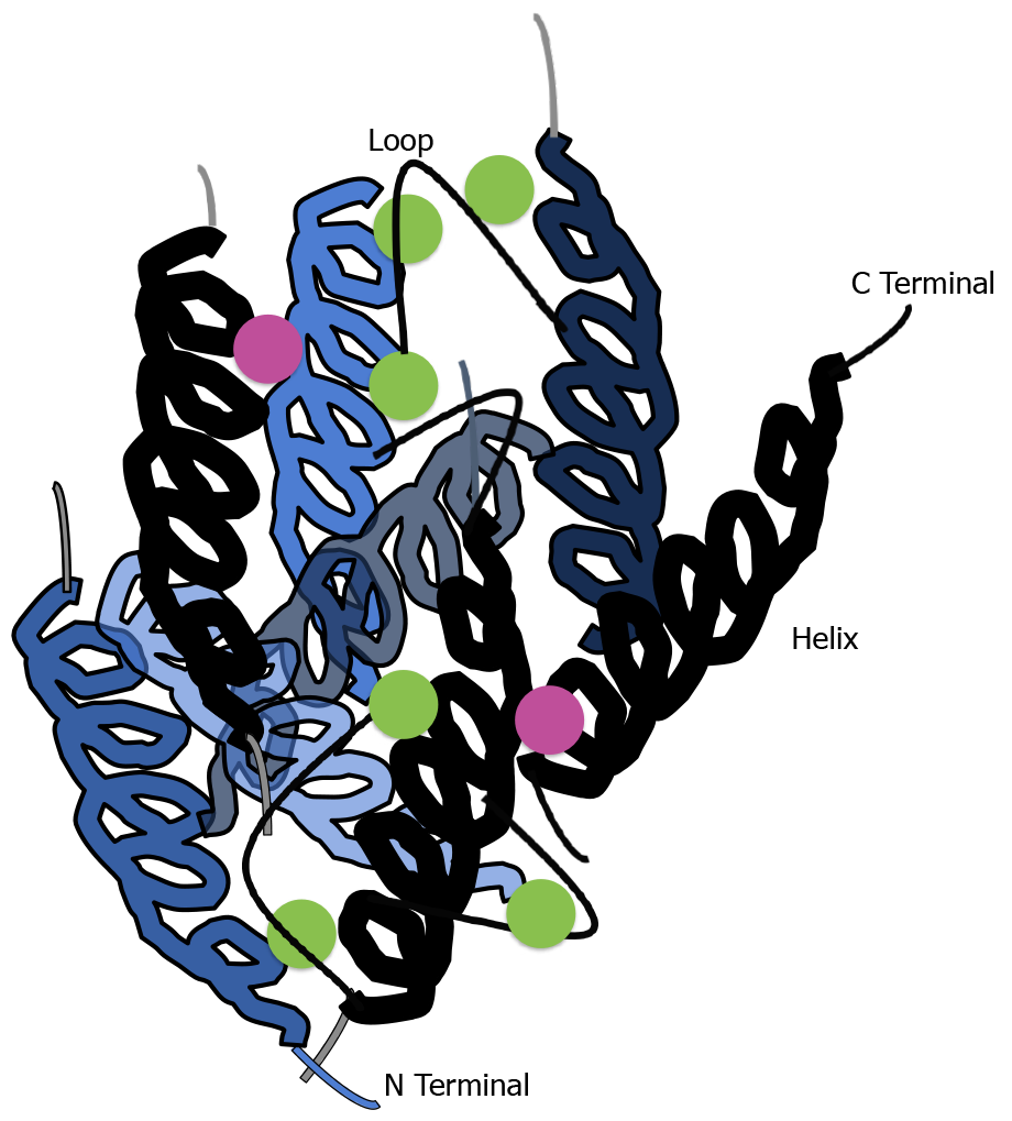
Figure 2 Diagrammatic representation of calprotectin dimer.
The crystal two S100A8-S100A9 Dimer structure of calprotectin loaded with Mn2+ and Ca2+. The black and blue chains represent S100A8 and S100A9, respectively. Purple spheres represent Mn2+ and green spheres represent Ca2+. Only one manganese ion can bind per calprotectin dimer.
- Citation: Al-Beltagi M, Saeed NK, Bediwy AS, Elbeltagi R. Fecal calprotectin in pediatric gastrointestinal diseases: Pros and cons. World J Clin Pediatr 2024; 13(2): 93341
- URL: https://www.wjgnet.com/2219-2808/full/v13/i2/93341.htm
- DOI: https://dx.doi.org/10.5409/wjcp.v13.i2.93341


