Copyright
©The Author(s) 2024.
World J Clin Pediatr. Jun 9, 2024; 13(2): 90641
Published online Jun 9, 2024. doi: 10.5409/wjcp.v13.i2.90641
Published online Jun 9, 2024. doi: 10.5409/wjcp.v13.i2.90641
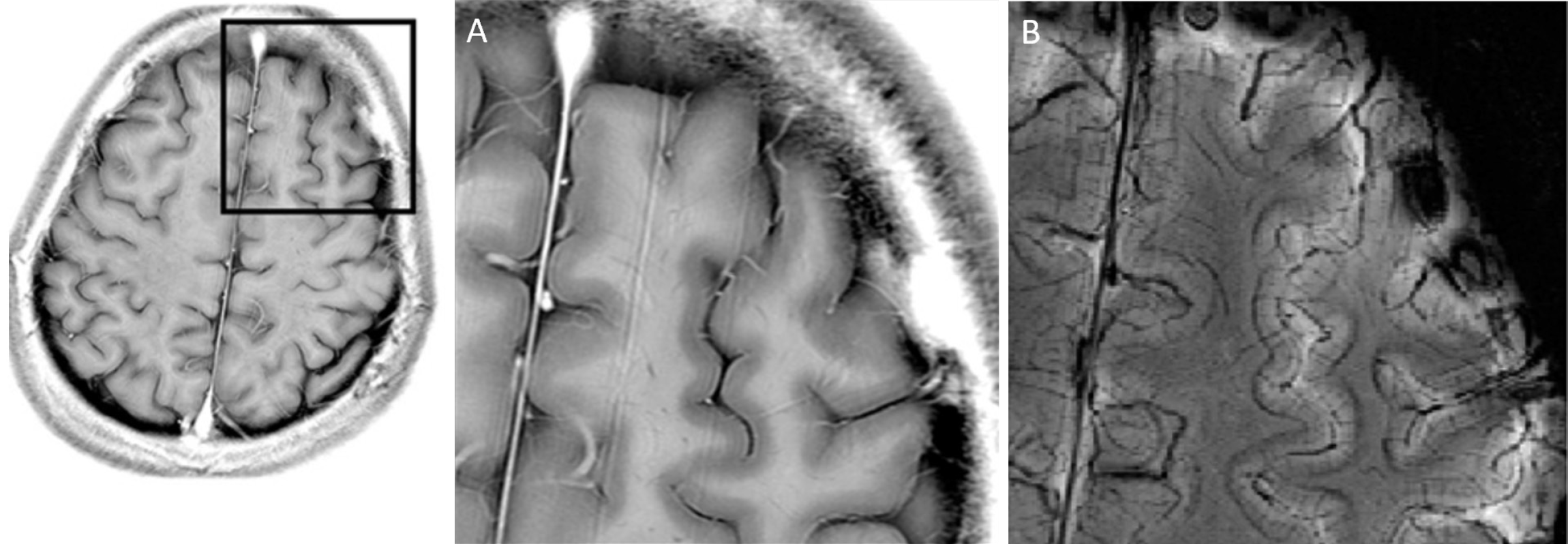
Figure 1 Comparative display of structural magnetic resonance images utilizing 3 Tesla and 7 Tesla systems.
A: Displaying an axial fast spin echo image and its magnified view in the left frontal region with the 3 Tesla system. The resolution is field of view (FOV) 200 mm/512 pixel = 0.39 mm/pixel, and the scan duration is approximately 5 min; B: Presenting an axial fast spin echo image and susceptibility-weighted image in the same slice using the 7 Tesla system. The resolution is FOV 80 mm/512 pixel = 0.16 mm/pixel, and the imaging time is approximately 4 min. A and B: Citation: Yamada K, Yoshimura J, Watanabe M, Suzuki K. Application of 7 tesla magnetic resonance imaging for pediatric neurological disorders: Early clinical experience. J Clin Imaging Sci 2021; 11: 65. Copyright© The Authors 2021. Published by Scientific Scholar. This is an open-access article distributed under the terms of the Creative Commons Attribution-Non Commercial-Share Alike 4.0 License, which allows others to remix, tweak, and build upon the work non-commercially, as long as the author is credited and the new creations are licensed under the identical terms. The authors have obtained the permission for figure using from Kenichi Yamada (Copyright permission).
- Citation: Perera Molligoda Arachchige AS, Politi LS. Potential applications of 7 Tesla magnetic resonance imaging in paediatric neuroimaging: Feasibility and challenges. World J Clin Pediatr 2024; 13(2): 90641
- URL: https://www.wjgnet.com/2219-2808/full/v13/i2/90641.htm
- DOI: https://dx.doi.org/10.5409/wjcp.v13.i2.90641


