Copyright
©The Author(s) 2024.
World J Clin Pediatr. Jun 9, 2024; 13(2): 90583
Published online Jun 9, 2024. doi: 10.5409/wjcp.v13.i2.90583
Published online Jun 9, 2024. doi: 10.5409/wjcp.v13.i2.90583
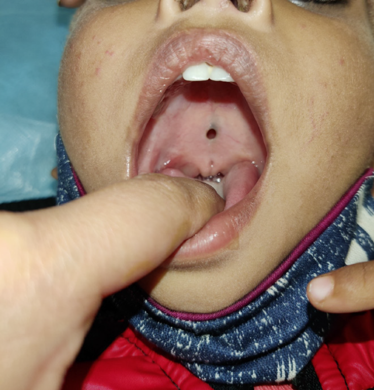
Figure 1 A submucous cleft palate is often missed until the child is much older and has poor speech development.
Note the zona pellucida and bifid uvula in this 3-year-old child.
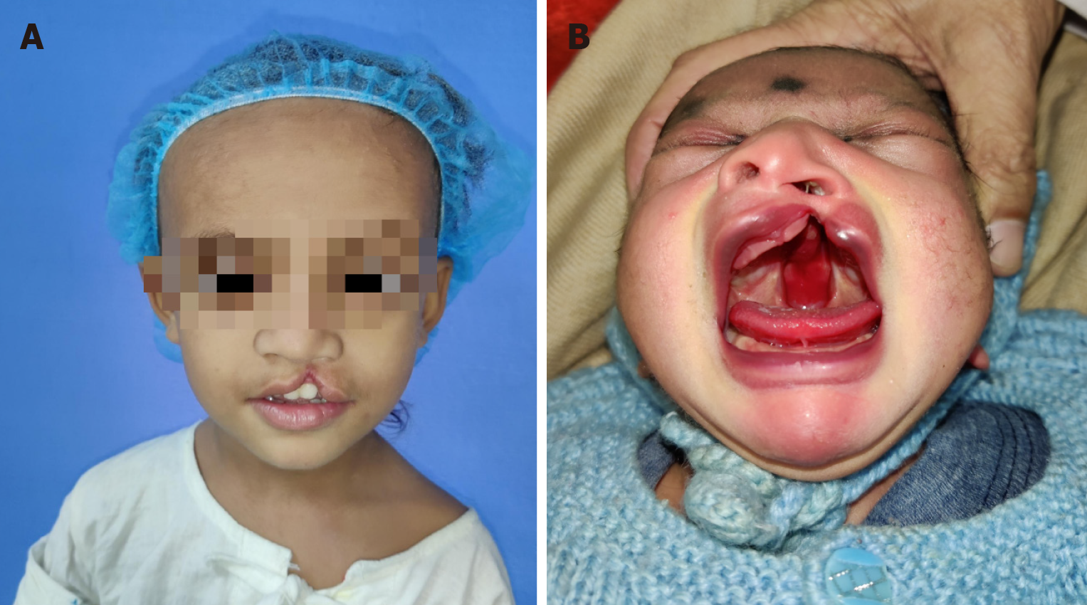
Figure 2 Cleft lip and palate.
A: 2-years-old boy with left unilateral incomplete cleft lip; B: Neonate with complete left cleft lip, alveolus and palate with Simonart band.
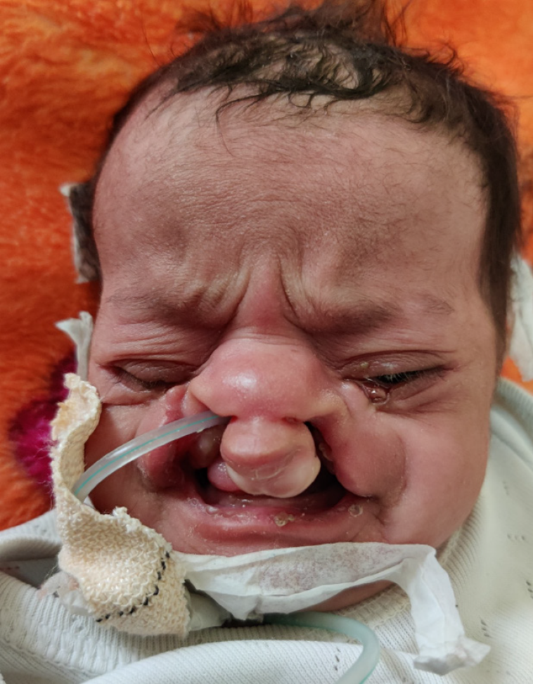
Figure 3
Neonate with a nasogastric tube inserted by the paediatrician for feeding.
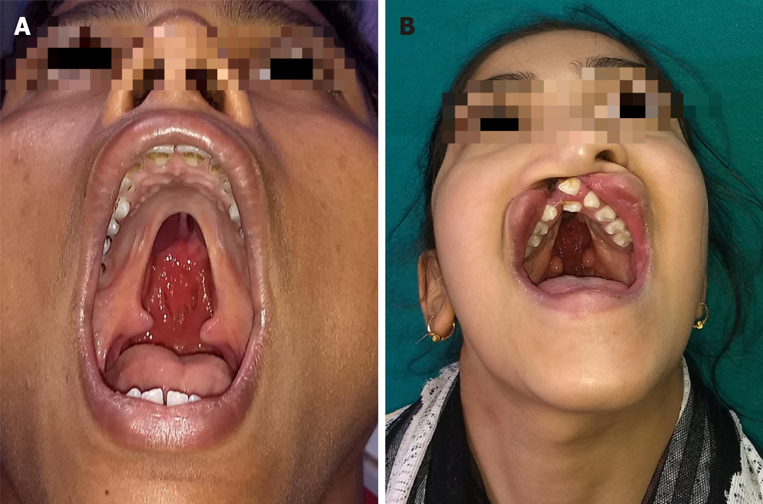
Figure 4 Cleft lip and palate in adults.
A: Normal maxillary growth in an adult un-operated cleft palate patient; B: Adult un-operated patient of cleft lip and palate with multiple dental abnormalities and nasal deformity.
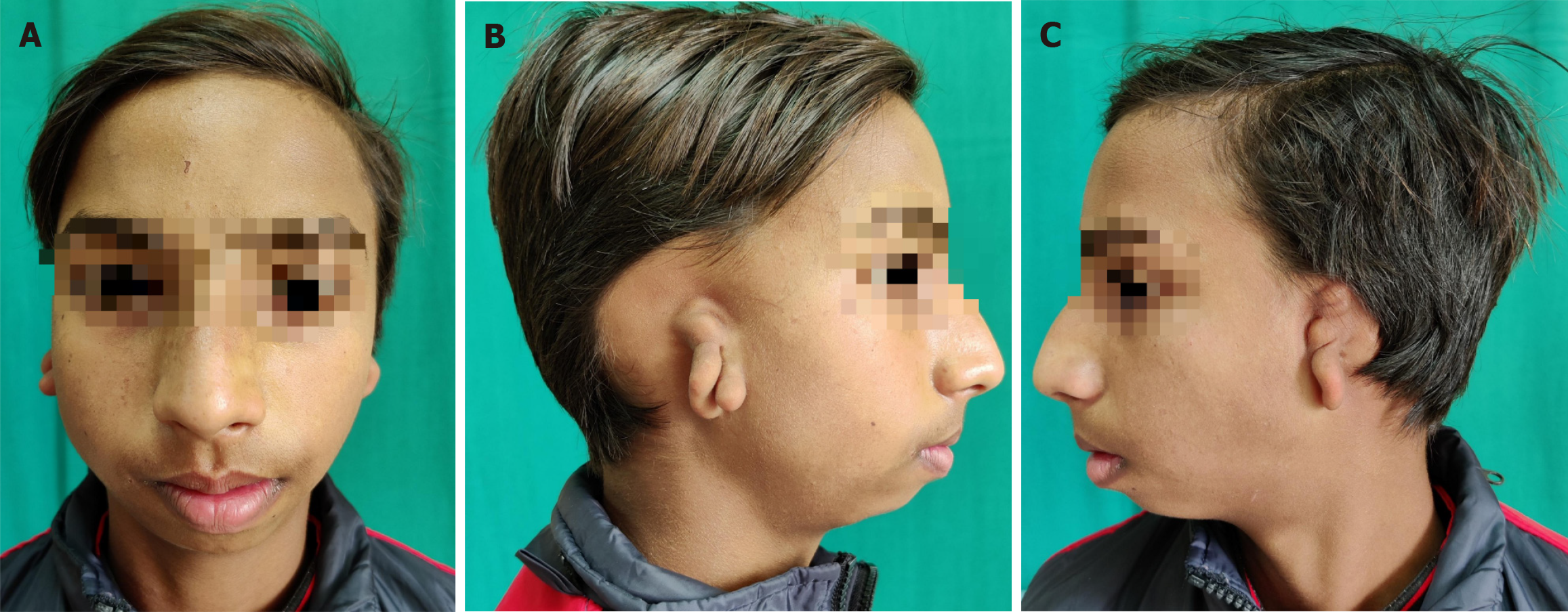
Figure 5 Bilateral microtia in a 12-year old boy.
A: Front view; B: Right ear; C: Left ear.
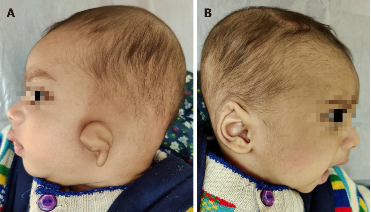
Figure 6 Neonate with unilateral lop ear.
A: Left affected ear; B: Right unaffected ear.
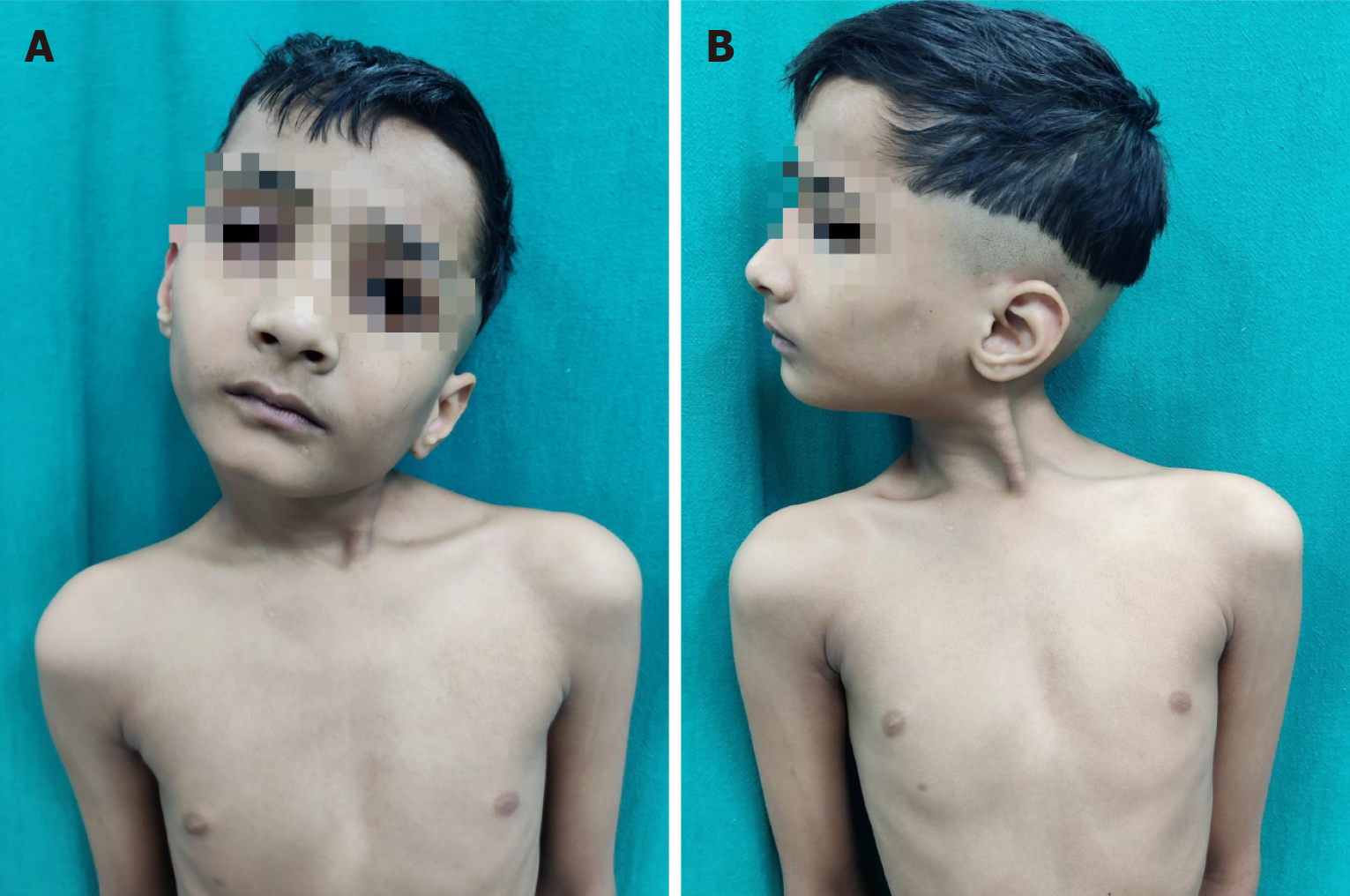
Figure 7 Eight-year-old child with left congenital muscular torticollis.
A: Note the shortened left sternocleidomastoid muscle as a fibrotic band, the facial asymmetry, restricted mobility of the neck; B: Lateral view of the same.
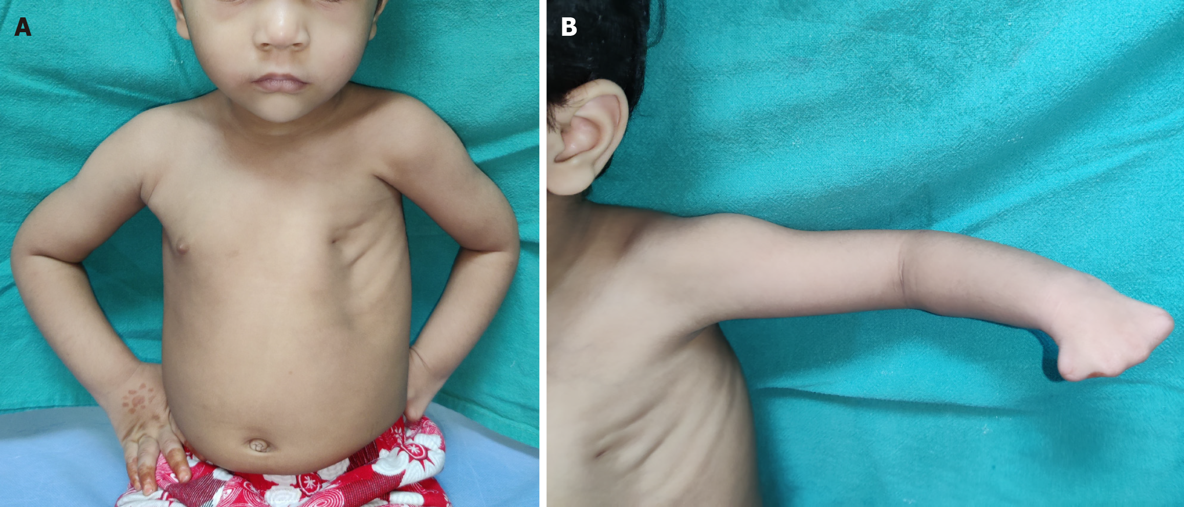
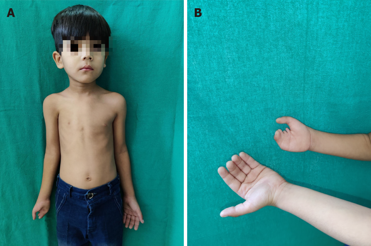
Figure 8 Poland syndrome in a 3-year-old girl child.
A: The chest can be reconstructed only after complete development of the opposite normal breast; B: Symbrachydactyly in the left hand.
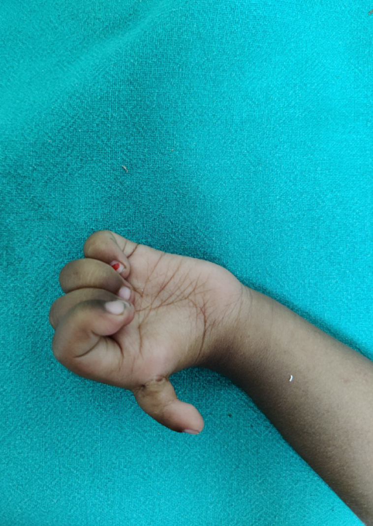
Figure 9 Poland syndrome in a 6-year-old male child brought to the out-patient department for right hand deformity.
A: The parents did not notice the mild hypoplastic right chest and nipple areola complex. He may not need or desire a chest reconstruction at all later in life; B: Right hand deformity.
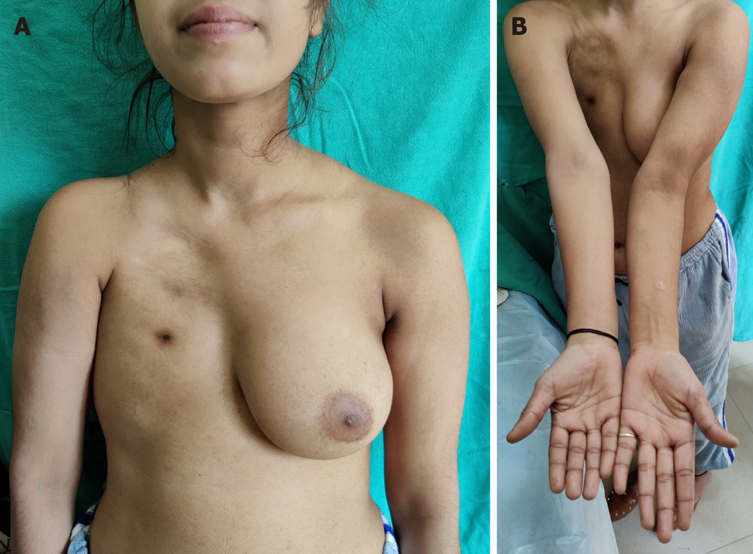
Figure 10 Poland syndrome in a girl in her late teens.
A: Right chest hypoplasia, absent breast seen. Reconstruction of right breast can be done with the left as a template; B: Short humerus on the affected side.
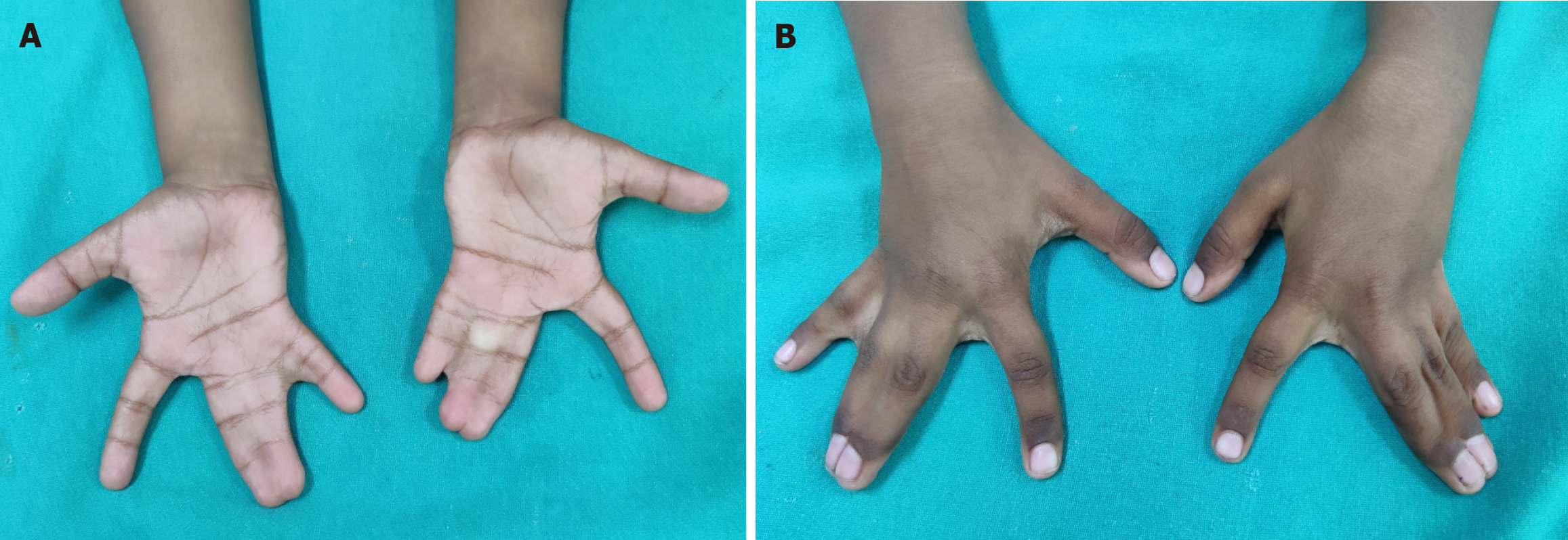
Figure 11 Syndactyly.
A: Complete syndactyly of bilateral 3rd web spaces and incomplete syndactyly 4th webspace (palmar view); B: Dorsal view.
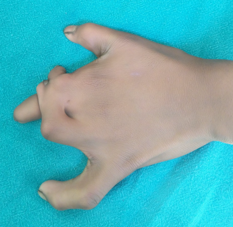
Figure 12
Complex complicated syndactyly with aberrant transverse bones and bony fusions.
Figure 13
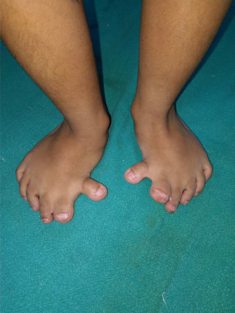
Bilateral feet pre-axial polydactyly and syndactyly in a teenager.
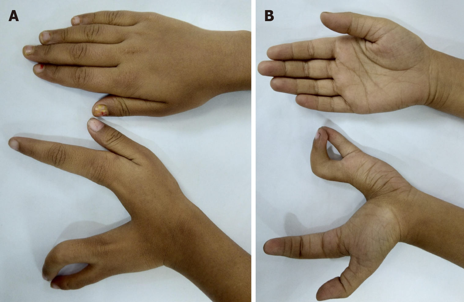
Figure 14 Left cleft hand with satisfactory function but poor aesthetic appearance.
A: Palmar view; B: Dorsal view.
Figure 15 ‘Pouce flottant’ thumb dysplasia should be operated with a correct balance between cortical plasticity and adequate size of tissues.
In many cases, if the thumb of the opposite side is normal, the child does not use the reconstructed thumb even after pollecization.
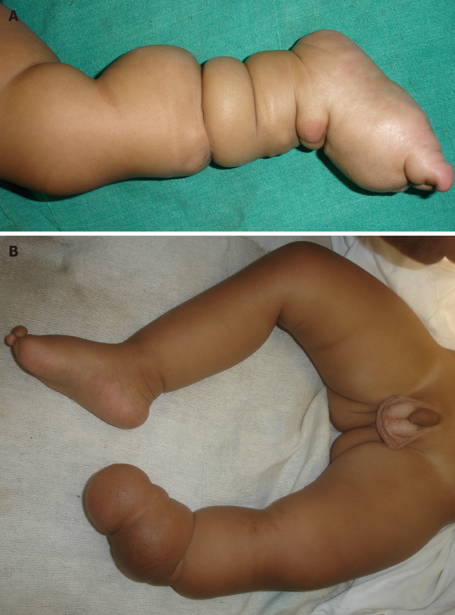
Figure 16 Constriction Ring Syndrome.
A: Multiple constriction rings in the lower limbs with lymphedema foot; B: Intrauterine amputation at level of distal left leg with proximal constriction rings seen.
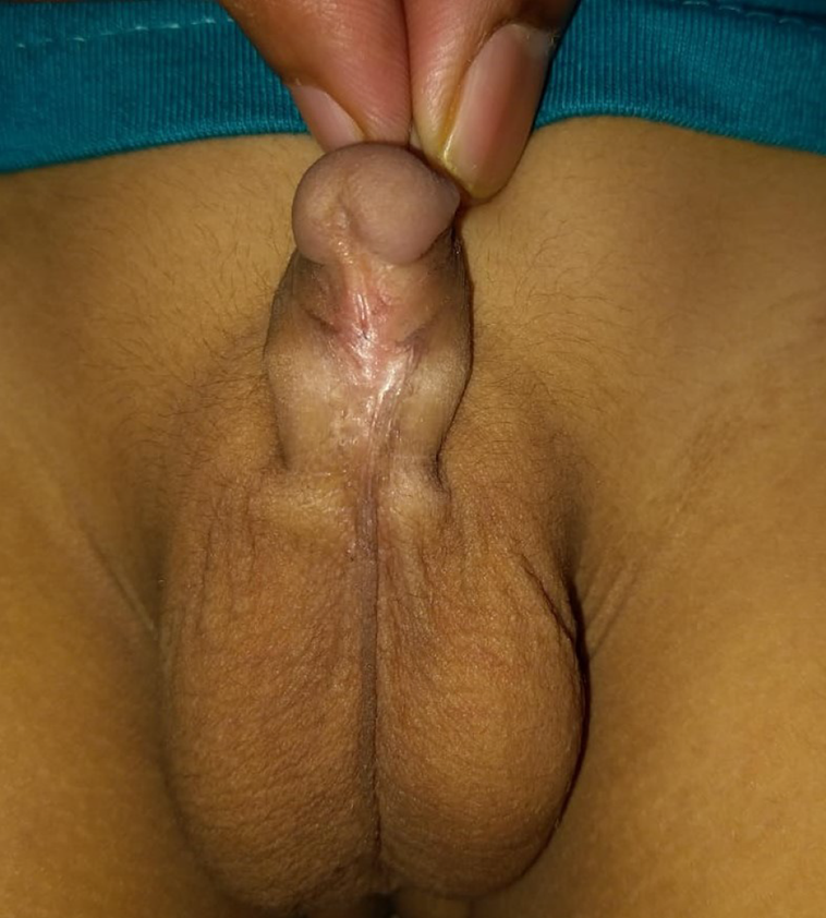
Figure 17
Mid penile hypospadias with chordee in an infant.
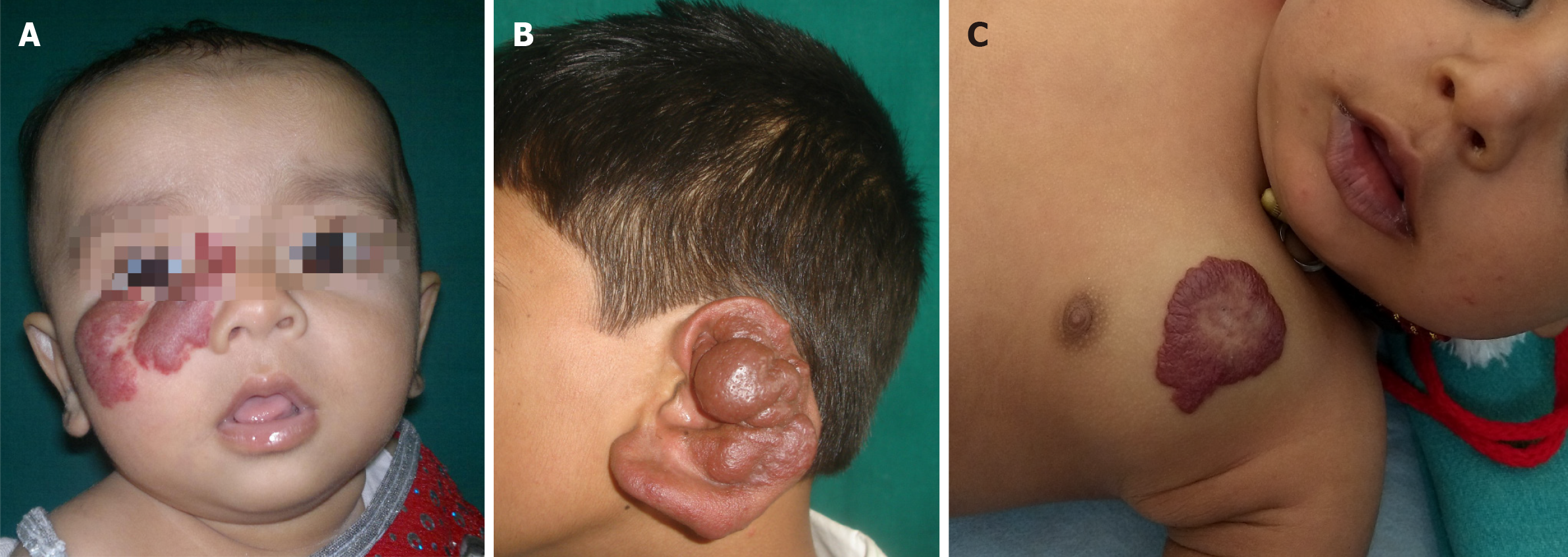
Figure 18 Vascular anomalies.
A: A hemangioma on the face of an infant. Proliferation of the lesion can cause obstruction to vision in the right eye, may need propranolol therapy to expedite involution; B: A pulsatile arterial malformation of the ear in a teenager, increasing in size- needs embolization and surgical intervention; C: An innocuous hemangioma on the chest- can follow the wait and watch policy for such a lesion.
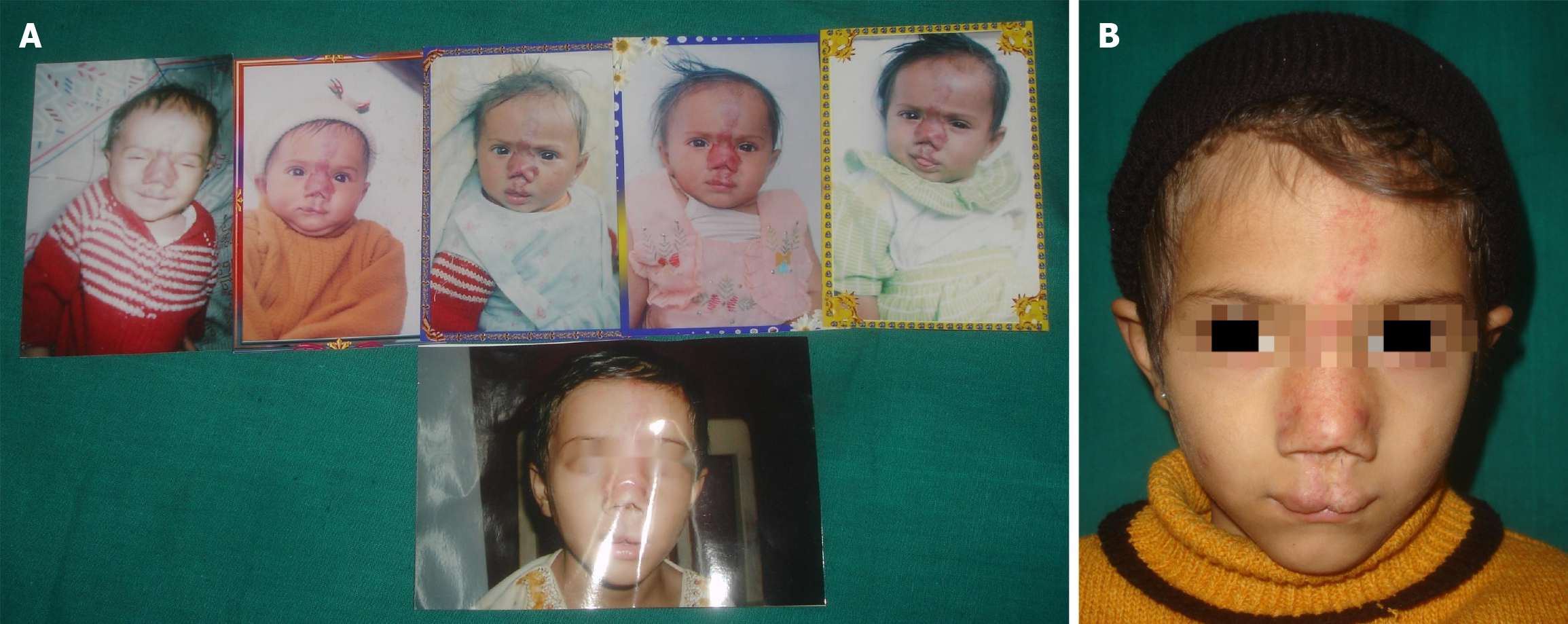
Figure 19 Congenital infantile hemagioma.
A: Serial photographic record of a child with vascular lesion on the face; B: Final photograph at 10 years of age with residual scarring.
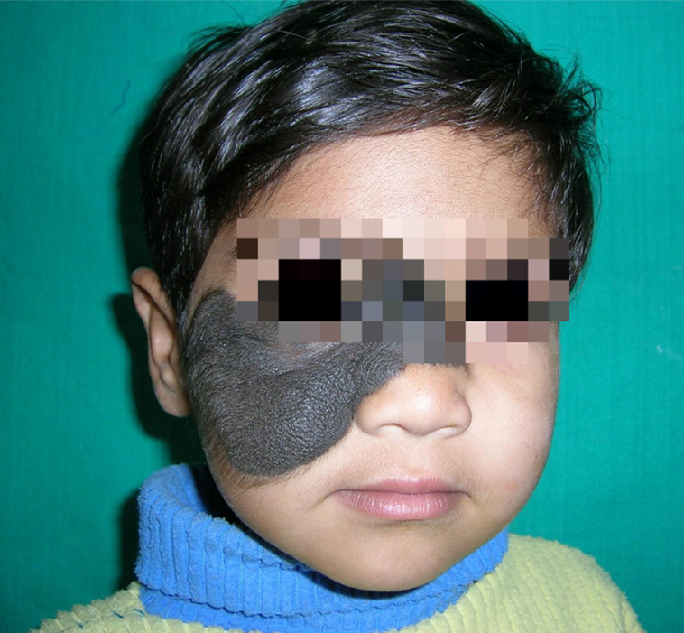
Figure 20
Congenital hairy nevus in a preschool toddler with a bear-like appearance- a huge stigma.
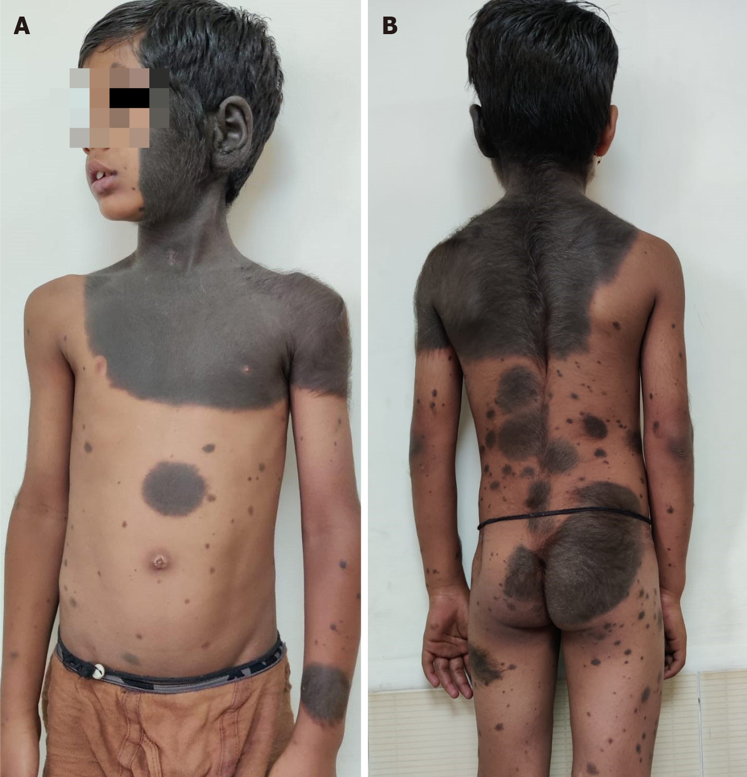
Figure 21 Giant hairy nevus.
A: A child with extensive involvement of the head and neck, trunk with multiple small and large satellite hairy nevi; B: Back, buttocks and limbs also have extensive involvement.
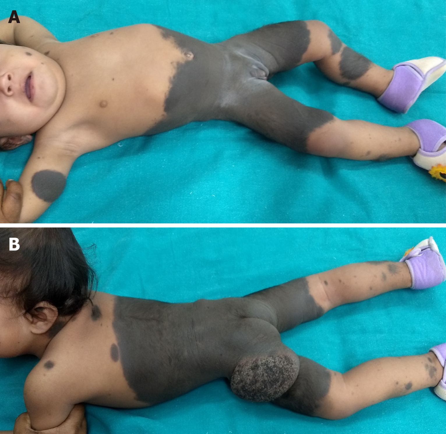
Figure 22 Bathing trunk nevus with satellite lesions.
There is not enough donor area for complete excision and resurfacing. A: Supine view; B: Prone view.
- Citation: Goel A, Goel A. Optimal timing for plastic surgical procedures for common congenital anomalies: A review. World J Clin Pediatr 2024; 13(2): 90583
- URL: https://www.wjgnet.com/2219-2808/full/v13/i2/90583.htm
- DOI: https://dx.doi.org/10.5409/wjcp.v13.i2.90583


