Copyright
©The Author(s) 2023.
World J Clin Pediatr. Jun 9, 2023; 12(3): 97-106
Published online Jun 9, 2023. doi: 10.5409/wjcp.v12.i3.97
Published online Jun 9, 2023. doi: 10.5409/wjcp.v12.i3.97
Figure 1 Computed tomography derived calcium score and role of angiography in convalescent phase of Kawasaki disease.
A: Computed tomography (CT) derived calcium scoring in a 19 years male patient on follow-up with history of Kawasaki disease in childhood shows a thickly calcified lesion in the proximal course of left anterior descending coronary artery (color marked as yellow with an arrow (calcium score was 910); B: Subsequent CT coronary angiographic curved reformatted image shows calcified aneurysm (arrow). LAD: Left anterior descending.
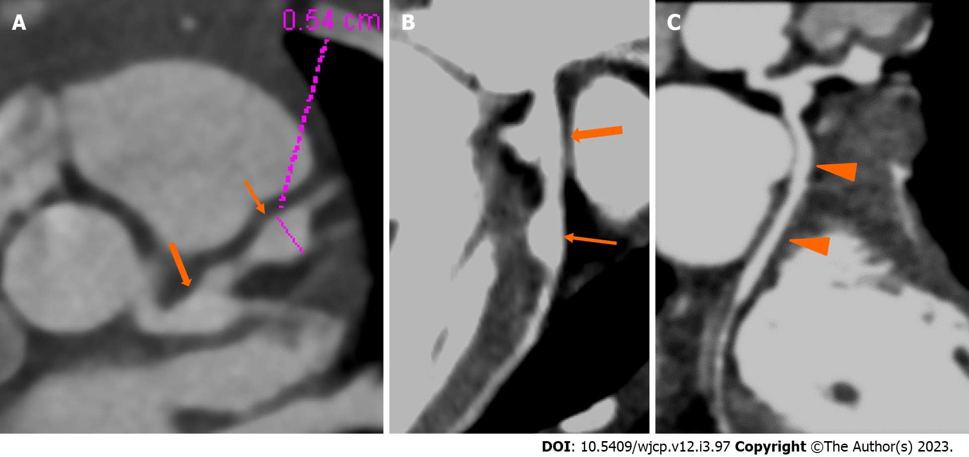
Figure 2 Computed tomography coronary angiography images showing its strength to evaluate coronary artery abnormalities in left circumflex artery.
A: 3 years male child at presentation shows fusiform aneurysm at bifurcation of left main coronary artery [left main coronary artery (LMCA)- thick arrows in A and B] with extension into osteo-proximal segment of left anterior descending (LAD). Note a skip fusiform aneurysm in proximal LAD (thin arrow in b). C: Proximal and mid segments of left circumflex (LCX) are dilated (arrow heads in C). Echo demonstrated LMCA and LAD aneurysms however cannot evaluate LCX due to its orientation and course.
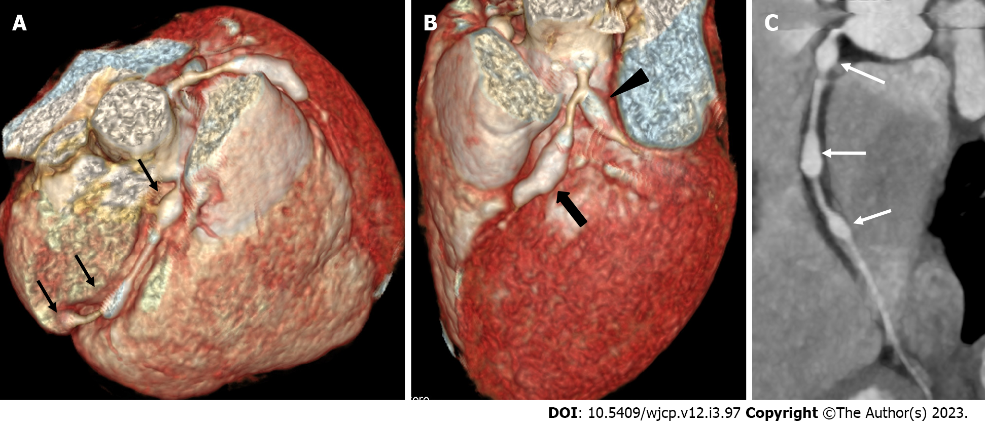
Figure 3 Computed tomography coronary angiography images showing its ability to evaluate mid and distal segments of coronary arteries.
Computed tomography coronary angiography (A and B: Volume rendered images; C: Curved reformatted image of RCA) of 4 years male at presentation demonstrate skip fusiform aneurysms in RCA (Thin arrows in A and C) and fusiform aneurysms in proximal LAD (thick arrow in B) and proximal left circumflex (arrow head in B). Fusiform aneurysm in mid and distal RCA could not be visualized on transthoracic echocardiography.
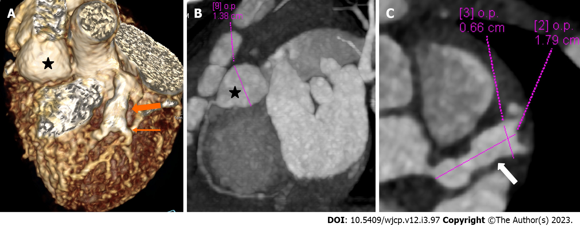
Figure 4 Panel of computed tomography coronary angiography images showing its ability to precisely identify location and morphology of aneurysms with extension into side branches.
Computed tomography coronary angiography (A: Volume rendered image; B: Curved reformatted image and C-axial image) of 4 years female child in acute phase (presentation) demonstrate giant saccular aneurysm in proximal resonance coronary angiography (asterisk in A and B). Fusiform aneurysm is seen in proximal left anterior descending (thick arrow in A) with extension into diagonal-1 branch (thin arrow in A).
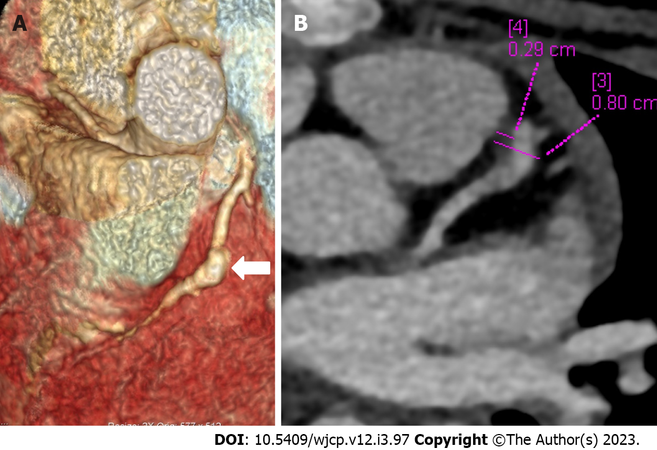
Figure 5 Computed tomography coronary angiography images depicting complications in coronary artery abnormalities (thrombus).
Computed tomography coronary angiography [(A: Volume rendered image; B: Axial image in plane of left anterior descending (LAD)] during follow up at shows a giant fusiform aneurysm in mid segment of LAD (thick arrow in A) with a hypodense plaque like tissue attached to the anterior wall (B) suggestive of thrombus. Child was having chest pain and ECHO at presentation and during the current episode was reported as normal; ECHO fails to elicit coronary artery abnormalities and its complication of thrombus in the aneurysm.
Figure 6 Computed tomography coronary angiography images showing its role in follow-up imaging and its role in assessment of compications.
Computed tomography coronary angiography (CTCA) at presentation in 1 year male (top row A-C) shows a giant complex aneurysm in mid segment of left anterior descending (LAD) (thick arrow in A and B) with multiple segmental aneurysms along the entire course of resonance coronary angiography (RCA). Follow-up CTCA after 3 years of presentation shows remodelling of LAD aneurysm (now becomes fusiform with mural calcification and severe stenosis its distal aspect (thick arrows D-F), also note resolution of aneurysmal dilatations of RCA (lower row D-F).
- Citation: Singhal M, Pilania RK, Gupta P, Johnson N, Singh S. Emerging role of computed tomography coronary angiography in evaluation of children with Kawasaki disease. World J Clin Pediatr 2023; 12(3): 97-106
- URL: https://www.wjgnet.com/2219-2808/full/v12/i3/97.htm
- DOI: https://dx.doi.org/10.5409/wjcp.v12.i3.97


