Copyright
©The Author(s) 2021.
World J Clin Pediatr. Mar 9, 2021; 10(2): 7-14
Published online Mar 9, 2021. doi: 10.5409/wjcp.v10.i2.7
Published online Mar 9, 2021. doi: 10.5409/wjcp.v10.i2.7
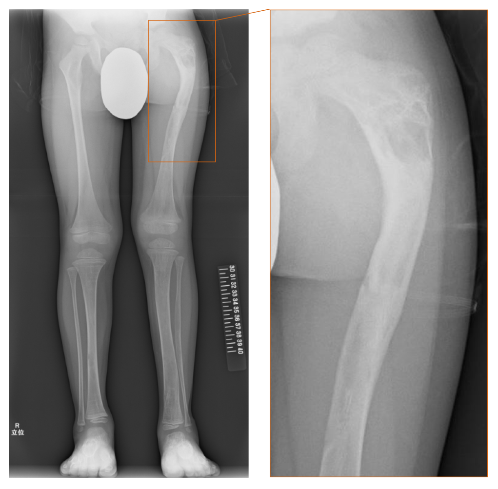
Figure 1 Radiograph at the age of 4 years and 8 mo.
The radiograph demonstrated a “ground-glass” appearance in his left femur and left tibia and “shepherd’s crook deformity” which is characterized by the presence of proximal femoral varus deformity and retroversion deformity, in his left thigh bone.
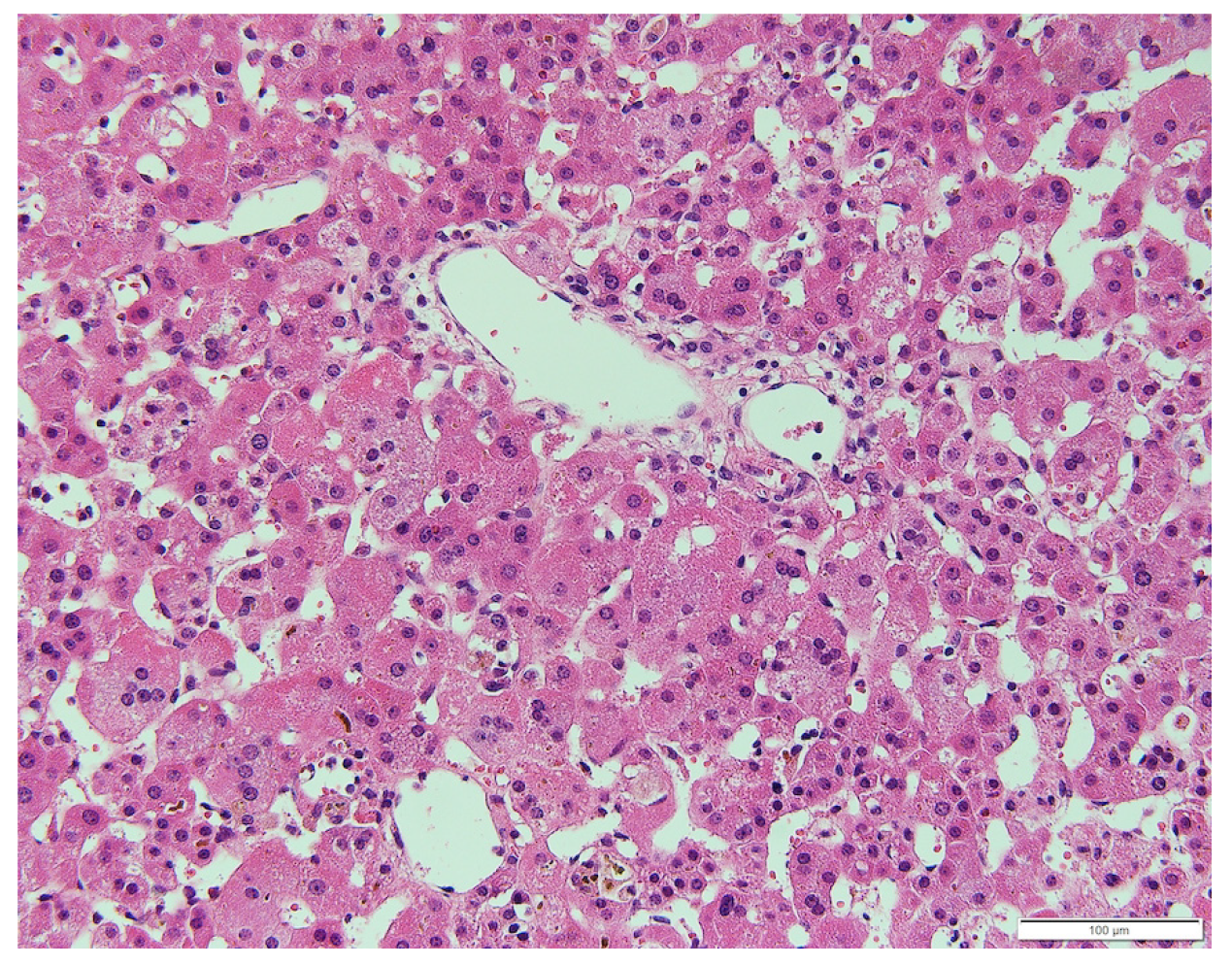
Figure 2 Liver specimen at the age of 1 mo.
Microscopic examination revealed a lack of bile ducts in the portal area and giant cell transformation of hepatocytes (hematoxylin and eosin staining).
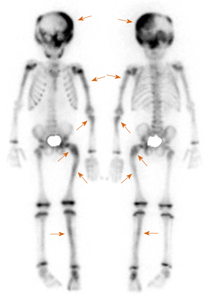
Figure 3 Bone scintigraphy with Tc-99 m-hydroxymethylene diphosphonate.
There are multiple hotspots with uptake at the left dominant skull and upper left limb in addition to the left femur and the left tibia.
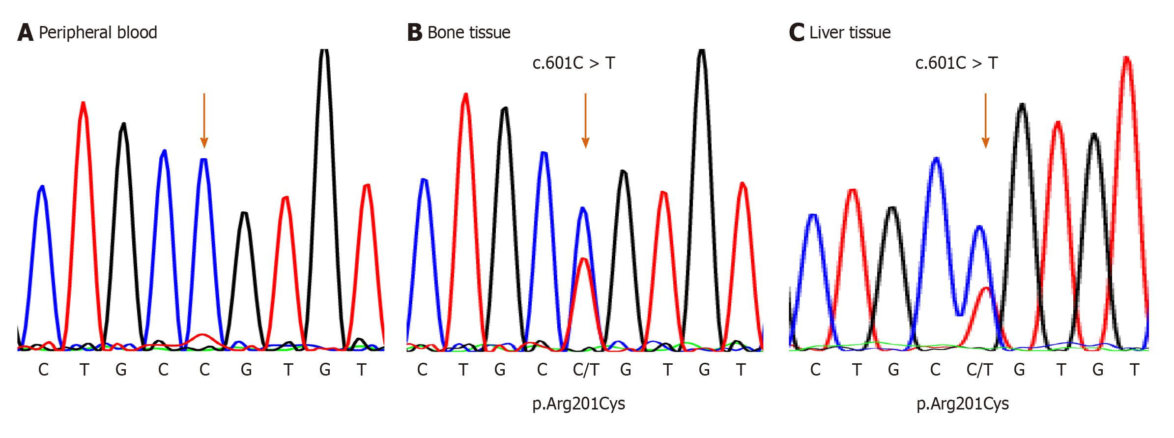
Figure 4 DNA sequencing of the GNAS gene.
A: Normal sequencing is shown in the peripheral blood; B and C: Arg201Cys mutation was detected in the bone tissue samples and the liver tissue.
- Citation: Satomura Y, Bessho K, Kitaoka T, Takeyari S, Ohata Y, Kubota T, Ozono K. Neonatal cholestasis can be the first symptom of McCune–Albright syndrome: A case report. World J Clin Pediatr 2021; 10(2): 7-14
- URL: https://www.wjgnet.com/2219-2808/full/v10/i2/7.htm
- DOI: https://dx.doi.org/10.5409/wjcp.v10.i2.7


