Published online May 20, 2015. doi: 10.5321/wjs.v4.i2.81
Peer-review started: September 26, 2014
First decision: December 17, 2014
Revised: January 7, 2015
Accepted: February 4, 2015
Article in press: February 6, 2015
Published online: May 20, 2015
Processing time: 238 Days and 18.9 Hours
Klippel-Feil syndrome (KFS) is defined by congenital cervical vertebral spine fusion and is seen with a wide spectrum of dental manifestations and craniofacial profiles. Previous studies on lateral cephalograms have documented an association between fusion of the cervical vertebrae and deviations in the craniofacial profile in non-syndromic patients with severe malocclusion. To our knowledge, no previous studies have described the craniofacial profile including the cranial base of KFS patients on lateral cephalograms. Therefore KFS and its craniofacial and dental manifestations were described according to existing literature and additionally the craniofacial profile and cranial base was analysed on lateral cephalograms of two patients with KFS. According to the literature the dental manifestations of KFS-patients included oligodontia, overjet, cross bite, open bite and deep bite. The craniofacial profile was clinically described as reduced lower facial height, midfacial hypoplasia, and mandibular prognathia. The analyses of the two lateral cephalograms showed increased mandibular inclination, increased vertical jaw-relationship, increased jaw angle and maxillary retrognathia. The cranial base was normal in both cases. The sagittal jaw relationship and mandibular prognathia varied between the two cases. The literature review and the analyses of the two lateral cephalograms have shown that deviations in the occipital and cervical spine field as KFS were associated with deviations in the teeth and craniofacial profile.
Core tip: Klippel-Feil syndrome (KFS) is defined by congenital cervical vertebral spine fusion and is seen with a wide spectrum of dental manifestations and craniofacial profiles. According to the literature dental manifestations of KFS-patients included oligodontia, horizontal maxillary overjet, cross bite, open bite and deep bite. The craniofacial profile was clinically described as reduced lower facial height, midfacial hypoplasia, and mandibular prognathia. Furthermore, two cases showed increased mandibular inclination, increased vertical jaw-relationship, increased jaw angle and maxillary retrognathia. The literature review and case analyses showed that deviations in the occipital and cervical spine field as KFS were associated with dentofacial deviations.
- Citation: Michelsen TG, Brusgaard PB, Sonnesen L. Klippel-feil: A syndrome in the occipital-cervical spine field and its dentofacial manifestations. World J Stomatol 2015; 4(2): 81-86
- URL: https://www.wjgnet.com/2218-6263/full/v4/i2/81.htm
- DOI: https://dx.doi.org/10.5321/wjs.v4.i2.81
Spranger et al[1] define syndromes accordingly: ”A syndrome is a pattern of multiple anomalies thought to be pathogenically related and not known to represent a single sequence or a polytopic field defect”. “Sequence” is defined as “A pattern of multiple anomalies derived from a single known or presumed prior anomaly or mechanical factor” and polytopic field defect is defined as “A pattern of anomalies derived from the disturbance of a single developmental field”[1]. Dorland’s Illustrated Medical Dictionary defines syndromes as: “A set of symptoms that occur together; the sum of signs of any morbid state; a symptom complex. In genetics, a pattern of multiple malformations thought to be pathogenically related”[2].
In Gorlin’s Syndromes of the Head and Neck[3] states that: “Congenital malformations of the head and neck are common and most resolve spontaneously within the first few days of postnatal life”. In some cases these “congenital malformations” turns out to be a part of a syndrome.
There are many different syndromes of the head- and neck-region and these syndromes are characterized by various clinical manifestations[3]. The manifestations can be specific for one particular syndrome, but different syndromes can have common manifestations[3]. Clinical manifestations can be revealed as skeletal deviations, soft-tissue-deviations or a combination of both[3].
Klippel-Feil syndrome (KFS) is a syndrome of the head- and neck-region. The syndrome has originally been described in the 16th Century, but was not named until 1912 by Hennekam et al[3]. KFS is defined by faulty segmentation of two or more cervical vertebrae[3] resulting in the occurrence of fusion of two or more cervical vertebrae on head film radiographs as lateral cephalograms. KFS is characterized by a triad of clinical symptoms such as short neck, limitation of head movement, and low posterior hairline. Furthermore, there are many associated anomalies in the craniofacial field in patients with KFS[3].
KFS is located in the occipital and cervical spine developmental field. The occipital and cervical spine field consists of structures of common embryological origin initiated by notochordal induction of the sclerotome formation in the somites, which develop into the cervical spine and the osseous structures in the occipital region[4-9]. The developmental field is funnel-shaped and limited anteriorly by the vertebral bodies, the basilar part of the occipital bone, and the postsphenoid bone. Posteriorly, the developmental field is limited by the cartilaginous part of the occipital bone and the vertebral arches[5,6].
A series of recent studies of non-syndromic patients with severe skeletal malocclusion traits have described the occurrence of cervical vertebral column fusion anomalies and analyzed the association between fusion anomalies and the craniofacial profile on lateral cephalograms[10-13]. These studies have documented significant associations between fusion and a large cranial base angle, between fusion and retrognathia of the jaws, and between fusion and inclination of the jaws. These findings indicate an association between fusion of the cervical vertebral column and the craniofacial profile including the cranial base in non-syndromic patients with severe skeletal malocclusion traits[10-14].
To our knowledge no previous studies have described the craniofacial profile including the cranial base of KFS patients on lateral cephalograms.
Therefore, the aim of the present study was to describe KFS and its craniofacial and dental manifestations according to previous literature. Additionally, the aim was to describe the craniofacial profile including the cranial base on lateral cephalograms of two patients with KFS.
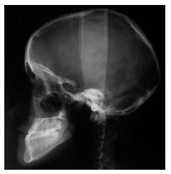
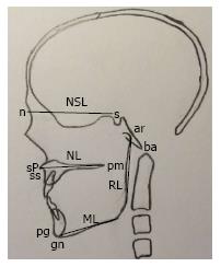
A literature review was performed in order to describe KFS and the dentofacial manifestation. Furthermore, lateral cephalograms of two patients with KFS with no other known symptoms (one boy, 8 years old and one girl, 15 years old; Figures 1 and 2) were included in the study. The two KFS patients comprise all KFS patients with no other known symptoms from Professor Sven Kreiborg’s archive, Department of Odontology, Copenhagen University. The craniofacial profile including the cranial base were measured by points and lines according to Solow and Tallgren[9]; illustrated in Figure 3. The landmarks used in the present study were marked on acetate sheets fixed to the radiograph. The variables were measured using a protractor and are shown in Table 1. The variables were compared to normal values of the craniofacial profile according to Björk et al[15] (Table 1). As the present study was a literature review and description of two lateral cephalograms no statistical analyses have been applied.
| Boy 8 yr(Figure 1)Case I | Girl 15 yr(Figure 2)Case II | Normal values according to Björk et al[15] | |
| Sagittal dimensions | |||
| s-n-ss | 78.5º | 76º | 82º (SD 3.5º) |
| s-n-pg | 75º | 86º | 80º (SD 3.5º) |
| ss-n-pg | 3.5º | -10º | 2º (SD 2.5º) |
| Vertical dimensions | |||
| NSL/NL | 9.5º | 8º | 8º (SD 3º) |
| NSL/ML | 39.5º | 37º | 33º (SD 6º) |
| NL/ML | 30º | 29º | 25º (SD 6º) |
| Cranial base | |||
| n-s-ba | 132º | 134º | 131º (SD 4.5º) |
| Jaw angle | |||
| ML/RL | 142º | 131º | 126º (SD 6º) |
KFS is a rare, congenital, skeletal malformation. It is defined by failure of normal segmentation of any two of the seven cervical vertebrae[16,17]. KFS may include fusion caudally of the cervical region. However, fusion in the lower spine, in the absence of cervical vertebral fusion, is not classified as KFS[18]. Generally, the second and the third vertebrae (C2-3)[18-20] and the fifth and sixth vertebrae (C5-6)[19] interspaces are most commonly fused. The C2-3 interspace fusion is thought to be an autosomal dominant inheritance, while C5-6 interspace fusion is considered to be autosomal recessive[19].
The absence of population screening studies has made it impossible to define the exact incidence and prevalence of KFS, but it has been estimated that it occurs in approximately 1:40.000-42.000 births[21,22]. Other studies have suggested, that KFS has an incidence of up to 0.5% of live births[18]. The incidence was none significantly slightly higher in females[19,21,22].
Although affected patients have cervical anomalies at birth, KFS is usually diagnosed at a later age[22]. It has been suggested that the fusion process in KFS patients is not fully present at birth and could be ongoing until skeletal maturity[20]. The disorder is often discovered incidentally when radiographs have been taken for other reasons[22]. The prognosis for most individuals diagnosed with KFS is good if the disorder is diagnosed early. But diagnostics of KFS is often complicated because the presence of cervical fusion cannot be determined in children younger than 8 years due to the development and ossification of the cervical vertebrae[17,22].
Occipitalization is also seen in KFS[20], and patients with atlantoaxial fusions are often diagnosed with KFS at younger ages than patients with more caudal fusions[22].
KFS is both affected by genetic and environmental factors and is morphologically and etiologically heterogeneous[3,23]. The heterogeneity of patients with KFS has made the diagnosis and classification difficult and has complicated elucidation of the genetic etiology of the syndrome[22]. Mutations in Pax1 have been found in patients with KFS, but the significance remains uncertain[3].
The earliest classification of KFS, by Feil, was based only on the anatomic distribution of the fused segments. Patients with KFS were assigned to one of three types[19]: Type I applied to patients with extensive cervical and upper thoracic fusion. Type II defined patients with one or two cervical interspace fusions and are often associated with hemivertebrae and occipitoatlantal fusion. Type III classified individuals with both cervical and lower thoracic or lumbar fusion[19].
Whereas Feils classification was based on the extent of vertebral fusion, a classification made by Clarke et al[18] focused on the etiology and genetic origins of the syndrome. Clarke et al[18] used three families, all affected with KFS, as a model for a new and comprehensive classification consisting of four different classes of KFS (KF1-4). Their study showed that an association exists between the position of the most rostral fusion, mode of inheritance, and some specific KFS associated anomalies[18].
Fusion of the cervical vertebrae can be symptomatic or asymptomatic. Some studies have found that up to 68% of KFS patients reported symptoms related to their syndrome[20]. The classic triad of KFS included low posterior hairline, short neck, and limitation of the neck movement[17,19]. However, the triad is present in only 50% of the patients[16,19,22]. Several other anomalies have been associated with KFS in varying degrees[18]. The anomalies included both systemic manifestations and craniofacial manifestations[24].
The most common manifestations in the craniofacial field are cleft palate[19,24], bifid uvula, and facial asymmetry[24]. Less frequently reported is the incidence of craniosynostosis and facial appearance. Facial appearance includes: reduced lower facial height, midfacial hypoplasia, mandibular malformation, and hypoplasia[19,24] and mandibular prognathia[25].
Moreover, KFS-patients show jaw anomalies, e.g., multiple jaw cysts, abnormal bony masses, duplication of the rami of the mandibular, and pseudoankylosis of the TMJ[24]. The documentation of the manifestations was based on clinical examinations and visual assessment of lateral cephalograms without any linear or angular measurements reported to describe the craniofacial profile.
The described dental manifestations included: oligodontia[17,24,26], horizontal maxillary overjet, cross bite, anterior open bite[24], and deep bite[17]. A case report has shown persistent primary teeth due to late eruption of the permanent dentition. Furthermore, the report showed velopharyngeal insufficiency causing difficulty in chewing and talking[17].
It has not been determined whether the craniofacial and dental findings were of random association or if they were truly related by any malformation mechanism of KFS[17].
The results of the analyses of the craniofacial profile on lateral cephalograms and the normal values according to Björk et al[15] are shown in Table 1.
Regarding the vertical dimensions of the craniofacial profile the KFS patients showed an increased mandibular inclination (NSL/ML), increased vertical jaw-relationship (NL/ML) and an increased jaw angle (ML/RL) compared to normal values (Table 1, Figures 1 and 2). In the sagittal plane the two cases showed retrognathia of the maxilla (s-n-ss) compared to the normal values whereas the prognathia of the mandible (s-n-pg) was larger in case II and smaller in case I. Furthermore, the sagittal jaw relationship (ss-n-pg) was larger in case I and smaller in case II compared to normal values (Table 1, Figures 1 and 2). The inclination of the maxilla (NSL/NL) and the cranial base angle (n-s-ba) was comparable to normal values.
KFS is a rare, congenital malformation defined by faulty segmentation of two or more cervical vertebrae[3]. Therefore, KFS is located in the occipital and cervical spine field[4-9]. The syndrome is morphologically and etiologically heterogeneous and within the group of KFS patients several anomalies have been reported[22]. In the literature the dental manifestations in KFS-patients were reported as oligodontia, horizontal maxillary overjet, cross bite, anterior open bite, and deep bite[24]. The craniofacial profile was clinically described as reduced lower face height, midface hypoplasia, and mandibular prognathia[24].
When comparing the clinical reports in the literature on the craniofacial profile with the two cases analyzed on lateral cephalograms in the present study some similarities are evident. The midface hypoplasia is described in the literature[24] and is also found in the two cases as retrognathia of the maxilla. Only in one case (case II) was mandibular prognathia seen in agreement with the clinical reports in the literature[24]. The increased inclination of the mandible, the increased vertical jaw-relationship, and the increased jaw angle indicating an increased lower face height in both cases in the present study was in disagreement with previous clinical reports in the literature[24]. The agreement and disagreements between the literature and the cases in the present study may reflect the morphologically and etiologically heterogeneous within the group of KFS patients[22] which complicates the understanding of this developmental syndrome[18].
Recently, a series of studies have shown significant associations between fusion of the cervical vertebrae and retrognathia of the jaws, between fusion and inclination of the jaws, and between fusion and a large cranial base angle in non-syndromic patients with severe skeletal malocclusion traits[10-12,14]. The measurements of the two KFS-cases showed both similar but also deviant patterns compared to those documented in the non-syndromic patients. Retrognathia of the maxilla was in agreement with previous findings in non-syndromic patients with fusion of the cervical vertebrae as well as inclination of the mandible[10-12,14]. On the other hand, retrognathia of the mandible was only seen in one case (case I) whereas mandibular prognathia was found in case II. Surprisingly, none of the cases showed a large cranial base angle, which was expected according to the literature[10-12,14].
An explanation for the association between the cervical spine and the craniofacial profile including the cranial base found in KFS and non-syndromic patients with fusion of the cervical vertebrae could be the notochord in the early embryogenesis[4]. The notochord develops in the human germ disc and determines the development of the cervical vertebrae, especially the vertebral bodies and the basilar part of the occipital bone in the cranial base (the posterior part of the cranial base angle)[4-9]. The para-axial mesoderm forming the vertebral arches and the remaining parts of the occipital bone are also formed from notochordal inductions[4-9]. The notochord is, by direct or indirect signaling, responsible for the formation of the structures in the occipital and cervical spine field in the early embryogenesis[4,5]. Therefore, a deviation in the development of the notochord may influence the surrounding bone tissue in the spine as well as in the posterior part of the cranial base to which the jaws are attached[10-14,27]. Furthermore, the jaws, including the condylar cartilage, develop from tissue that derives from the neural crest. The neural crest cells migrate to the craniofacial area before the notochord is surrounded by bone tissue and disappears[4]. In the first branchial arch, the neural crest cells migrate from the neural crest towards the mandible, followed by the cells to the maxilla, and lastly by the cells to the nasofrontal region[4] before the notochord is surrounded by bone tissue[28]. Therefore, it is understandable that a disturbance in the amount of migrating maxillary and mandibular cells or timing of the migration of the maxillary and mandibular cells may influence both the sagittal development (retrognathia of the jaws) and vertical development (inclination of the jaws)[11-13]. How the migration of the neural crest cells are influenced by signals from the notochord is still unclear, but signaling during early embryogenesis between the notochord, para-axial mesoderm, the neural tube, and the neural crest, as described above, is believed to be important for the associations between malformation of the craniofacial structures and the cervical vertebrae[14,27].
The associations found in KFS and in non-syndromic patients with severe malocclusion traits between fusion of the cervical vertebrae and deviations in the craniofacial profile may lead to considerations regarding etiology and classification of KFS.
KFS is defined by congenital vertebral fusion of the cervical spine and is seen with a wide spectrum of dental manifestations and craniofacial profile. According to the literature the dental manifestations of KFS-patients include oligodontia, horizontal maxillary overjet, cross bite, open bite and deep bite. The craniofacial profile is clinically described as reduced lower facial height, midfacial hypoplasia, and mandibular prognathia.
The analyses of the two lateral cephalograms showed increased mandibular inclination, increased vertical jaw-relationship, increased jaw angle and maxillary retrognathia. The cranial base was normal in both cases. The sagittal jaw relationship and mandibular prognathia varied between the two cases.
The literature review and the analyses of the two lateral cephalograms have shown that deviations in the occipital and cervical spine field as KFS were associated with deviations in the teeth and craniofacial profile.
Professor Sven Kreiborg is acknowledged for providing the two lateral cephalograms from his archive; and Maria Kvetny for linguistic support and Gyldan Zejnelovska for manuscript preparation.
P- Reviewer: Teli MGA, Tokuhashi Y, Zhou M S- Editor: Ji FF L- Editor: A E- Editor: Jiao XK
| 1. | Spranger J, Benirschke K, Hall JG, Lenz W, Lowry RB, Opitz JM, Pinsky L, Schwarzacher HG, Smith DW. Errors of morphogenesis: concepts and terms. Recommendations of an international working group. J Pediatr. 1982;100:160-165. [PubMed] |
| 2. | Dorland , W A Newman. Dorland’s Illustrated Medical Dictionary. 30th ed. United States, Philadelphia, PA: Saunders 2003; . |
| 3. | Hennekam RCM, Krantz ID, Allanson JE. Gorlin’s syndromes of the head and neck. 5th ed. New York: Oxford University Press 2010; . |
| 4. | Kjaer I. Neuro-osteology. Crit Rev Oral Biol Med. 1998;9:224-244. [PubMed] |
| 5. | Kjaer I. Orthodontics and foetal pathology: a personal view on craniofacial patterning. Eur J Orthod. 2010;32:140-147. [RCA] [PubMed] [DOI] [Full Text] [Cited by in Crossref: 36] [Cited by in RCA: 37] [Article Influence: 2.3] [Reference Citation Analysis (0)] |
| 6. | Kjær I. Dental approach to craniofacial syndromes: how can developmental fields show us a new way to understand pathogenesis? Int J Dent. 2012;2012:145749. [RCA] [PubMed] [DOI] [Full Text] [Full Text (PDF)] [Cited by in Crossref: 10] [Cited by in RCA: 9] [Article Influence: 0.7] [Reference Citation Analysis (0)] |
| 7. | Müller F, O’Rahilly R. The early development of the nervous system in staged insectivore and primate embryos. J Comp Neurol. 1980;193:741-751. [PubMed] |
| 8. | Sadler TW. Embryology of neural tube development. Am J Med Genet C Semin Med Genet. 2005;135C:2-8. [PubMed] |
| 9. | Solow B, Tallgren A. Head posture and craniofacial morphology. Am J Phys Anthropol. 1976;44:417-435. [PubMed] |
| 10. | Sonnesen L, Kjaer I. Cervical vertebral body fusions in patients with skeletal deep bite. Eur J Orthod. 2007;29:464-470. [PubMed] |
| 11. | Sonnesen L, Kjaer I. Cervical column morphology in patients with skeletal Class III malocclusion and mandibular overjet. Am J Orthod Dentofacial Orthop. 2007;132:427.e7-427.12. [PubMed] |
| 12. | Sonnesen L, Kjaer I. Anomalies of the cervical vertebrae in patients with skeletal Class II malocclusion and horizontal maxillary overjet. Am J Orthod Dentofacial Orthop. 2008;133:188.e15-20. [RCA] [DOI] [Full Text] [Cited by in Crossref: 35] [Cited by in RCA: 32] [Article Influence: 1.9] [Reference Citation Analysis (0)] |
| 13. | Sonnesen L, Kjaer I. Cervical column morphology in patients with skeletal open bite. Orthod Craniofac Res. 2008;11:17-23. [RCA] [PubMed] [DOI] [Full Text] [Cited by in Crossref: 40] [Cited by in RCA: 39] [Article Influence: 2.3] [Reference Citation Analysis (0)] |
| 14. | Sonnesen L. Associations between the Cervical Vertebral Column and Craniofacial Morphology. Int J Dent. 2010;2010:295728. [RCA] [PubMed] [DOI] [Full Text] [Full Text (PDF)] [Cited by in Crossref: 28] [Cited by in RCA: 26] [Article Influence: 1.7] [Reference Citation Analysis (0)] |
| 15. | Björk A, Hasund A, Jacobsen U, Lundström A, Myrberg N, Slagsvold O. Nordisk Lärebok i ortodonti. Stockholm: Sveriges Tandläkarförbunds Förlagsförening 1971; . |
| 16. | Auerbach JD, Hosalkar HS, Kusuma SK, Wills BP, Dormans JP, Drummond DS. Spinal cord dimensions in children with Klippel-Feil syndrome: a controlled, blinded radiographic analysis with implications for neurologic outcomes. Spine (Phila Pa 1976). 2008;33:1366-1371. [RCA] [PubMed] [DOI] [Full Text] [Cited by in Crossref: 15] [Cited by in RCA: 14] [Article Influence: 0.8] [Reference Citation Analysis (0)] |
| 17. | Barbosa V, Maganzini AL, Nieberg LG. Dento-skeletal implications of Klippel-Feil syndrome [a case report]. N Y State Dent J. 2005;71:48-51. [PubMed] |
| 18. | Clarke RA, Catalan G, Diwan AD, Kearsley JH. Heterogeneity in Klippel-Feil syndrome: a new classification. Pediatr Radiol. 1998;28:967-974. [PubMed] |
| 19. | Nagib MG, Maxwell RE, Chou SN. Klippel-Feil syndrome in children: clinical features and management. Childs Nerv Syst. 1985;1:255-263. [PubMed] |
| 20. | Samartzis D, Kalluri P, Herman J, Lubicky JP, Shen FH. Superior odontoid migration in the Klippel-Feil patient. Eur Spine J. 2007;16:1489-1497. [PubMed] |
| 21. | Kaplan KM, Spivak JM, Bendo JA. Embryology of the spine and associated congenital abnormalities. Spine J. 2005;5:564-576. [PubMed] |
| 22. | Tracy MR, Dormans JP, Kusumi K. Klippel-Feil syndrome: clinical features and current understanding of etiology. Clin Orthop Relat Res. 2004;424:183-190. [PubMed] |
| 23. | O’Rahilly R, Müller F. Human Embryology and Teratology. 3rd ed. USA: Wiley-Liss 2001; . |
| 24. | Naikmasur VG, Sattur AP, Kirty RN, Thakur AR. Type III Klippel-Feil syndrome: case report and review of associated craniofacial anomalies. Odontology. 2011;99:197-202. [RCA] [PubMed] [DOI] [Full Text] [Cited by in Crossref: 13] [Cited by in RCA: 15] [Article Influence: 1.1] [Reference Citation Analysis (0)] |
| 25. | Papagrigorakis MJ, Gisakis IG, Synodinos F, Fakitsas I. Klippel-Feil syndrome: report of a case with craniofacial complex involvement. Hellenic Orthodontic Rev. 2004;117-128. |
| 26. | Papagrigorakis MJ, Synodinos PN, Daliouris CP, Metaxotou C. De novo inv(2)(p12q34) associated with Klippel-Feil anomaly and hypodontia. Eur J Pediatr. 2003;162:594-597. [PubMed] |
| 27. | Sonnesen L, Pedersen CE, Kjaer I. Cervical column morphology related to head posture, cranial base angle, and condylar malformation. Eur J Orthod. 2007;29:398-403. [PubMed] |
| 28. | Gary CS, Steven BB, Philip RB, Philippa HF-W. Larsen’s Human Embryology. 4th ed. London: Churchill Livingstone 2009; . |


