Copyright
©The Author(s) 2019.
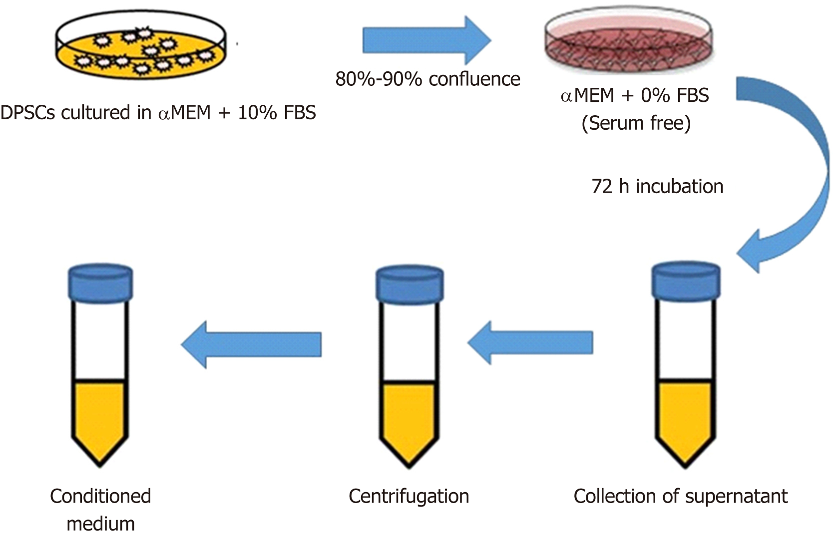
Figure 1 Schematic diagram representing the steps of obtaining cell-free conditioned medium from dental pulp stem cells.
The dental pulp stem cells (DPSCs) were plated onto culture vessels and grown in culture medium containing 10% fetal bovine serum. When adherent cells reach 80%-90% confluence, the medium was discarded and the cells washed with phosphate-buffered saline. Then, the DPSCs were incubated in α MEM serum-free medium and incubated for 72 h. The supernatant representing the conditioned medium was collected and centrifuged, filtered and concentrated.
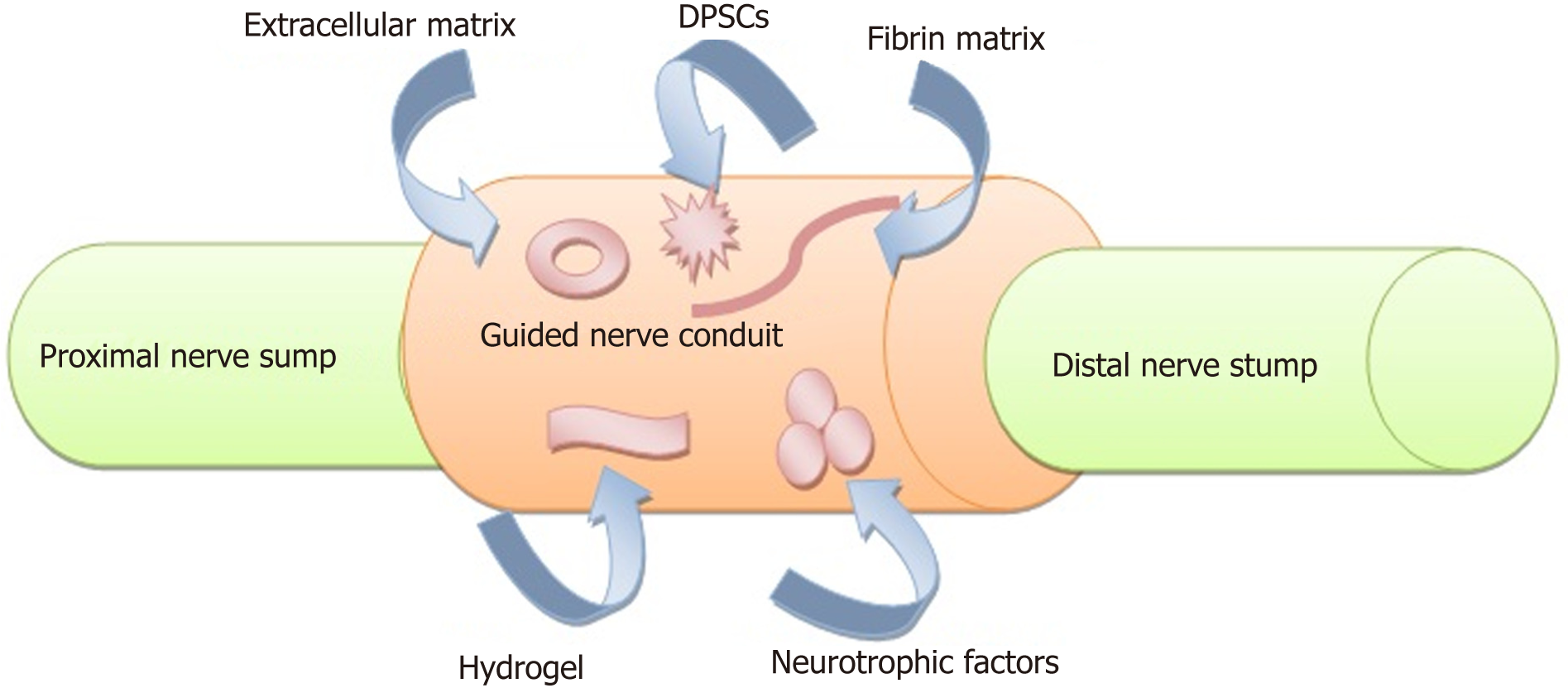
Figure 2 Schematic diagram showing a strategy to promote regeneration of bisection peripheral nerve injury using guided nerve conduit with incorporation of dental pulp stem cells and neurotrophic factors.
The proximal and distal nerve stumps (light green color) are sutured into the two ends of artificial nerve conduit (peach color). The conduit mimics the structures and components of autologous nerves and bridging the nerve gap to support the growth and regeneration of neural cells. The microenvironment of the conduit should contain nutrients, cytokines and growth factors (extracellular matrix, hydrogel and neurotrophic factors) as well as cellular elements (dental pulp stem cells).
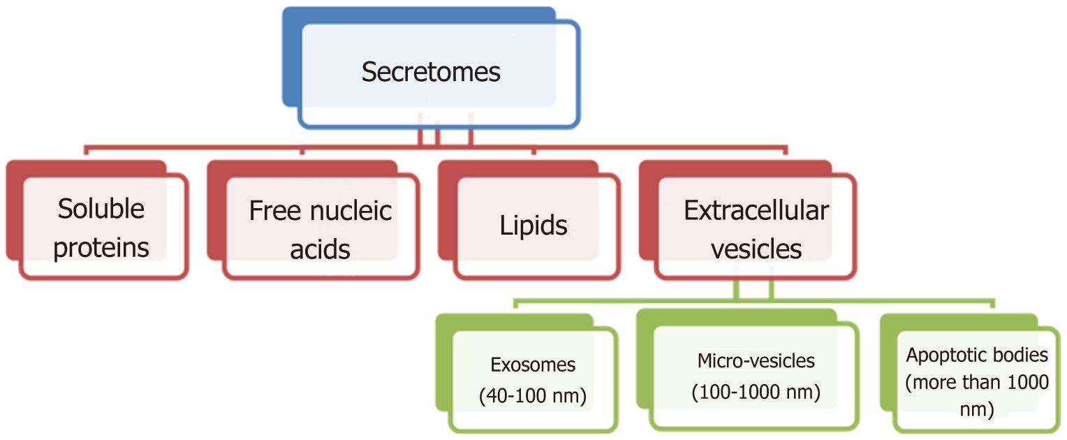
Figure 3 Organization chart representing the different components of secretomes from the mesenchymal stem cells.
Exosomes, micro-vesicles and apoptotic bodies were classified according to the size and morphology. Exosomes (40-100 nm) usually derived from multi-vesicular bodies, microvesicles (100-1000 nm) by plasma membrane budding and anti-apoptotic bodies (more than 1000 nm) via blebbing of plasma membrane of dying cells.
Figure 4 Horizontal bullet list for the dental pulp stem cells’ secretome, their mechanism of action and the involved factors in the immunomodulatory, anti-inflammatory, anti-apoptotic angiogenic regulatory and neurotrophic function.
- Citation: Sultan N, Amin LE, Zaher AR, Scheven BA, Grawish ME. Dental pulp stem cells: Novel cell-based and cell-free therapy for peripheral nerve repair. World J Stomatol 2019; 7(1): 1-19
- URL: https://www.wjgnet.com/2218-6263/full/v7/i1/1.htm
- DOI: https://dx.doi.org/10.5321/wjs.v7.i1.1


