Copyright
©2013 Baishideng Publishing Group Co.
World J Stomatol. Nov 20, 2013; 2(4): 86-90
Published online Nov 20, 2013. doi: 10.5321/wjs.v2.i4.86
Published online Nov 20, 2013. doi: 10.5321/wjs.v2.i4.86
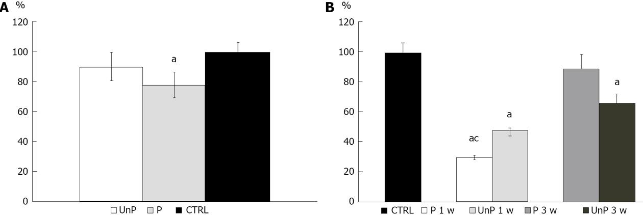
Figure 1 Histogram of cell viability.
A: Cell viability of fibroblast cultured directly on unpolished samples (UnP), polished samples (P: finished surface using polishing discs) and control (CTRL); B: Cell viability of fibroblasts in CTRL, polished samples at 1 wk (P 1 w), unpolished samples at 1 wk (UnP 1 w), polished samples at 3 wk (P 3 w), unpolished samples at 3 wk (UnP 3 w); aP < 0.05 vs CTRL; cP < 0.05 P 1 w vs P 3 w.
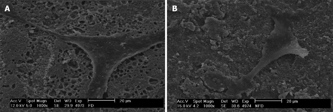
Figure 2 Scanning electron micrograph (x 2000, magnification).
A: Gingival fibroblasts cultured directly on polished sample; B: Gingival fibroblasts cultured directly on unpolished sample.
- Citation: Orsini G, Catellani A, Ferretti C, Gesi M, Mattioli-Belmonte M, Putignano A. Cytotoxicity of a silorane-based dental composite on human gingival fibroblasts. World J Stomatol 2013; 2(4): 86-90
- URL: https://www.wjgnet.com/2218-6263/full/v2/i4/86.htm
- DOI: https://dx.doi.org/10.5321/wjs.v2.i4.86


