Copyright
©2013 Baishideng Publishing Group Co.
World J Stomatol. Aug 20, 2013; 2(3): 62-66
Published online Aug 20, 2013. doi: 10.5321/wjs.v2.i3.62
Published online Aug 20, 2013. doi: 10.5321/wjs.v2.i3.62
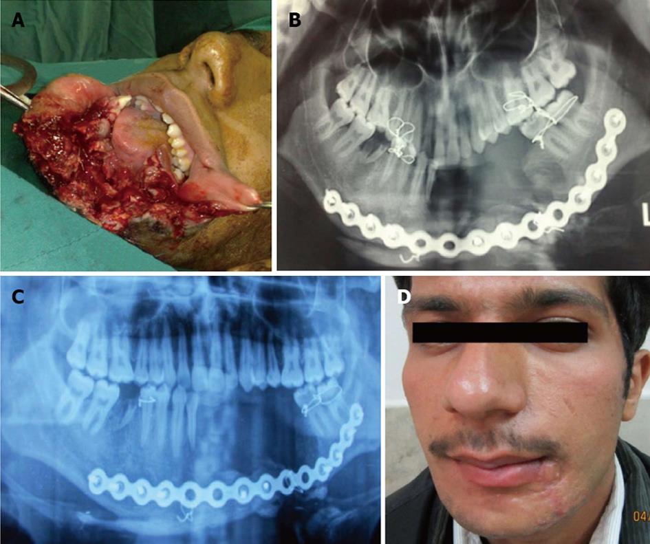
Figure 1 A 20-year old man with a gunshot wound to the lower face, with disruption of soft tissue and the mandible bone in body with bone defect.
A: Before operation; B: Computed tomography scan before operation; C: Reconstruction with reconstructive plate; D: Three months post operation.
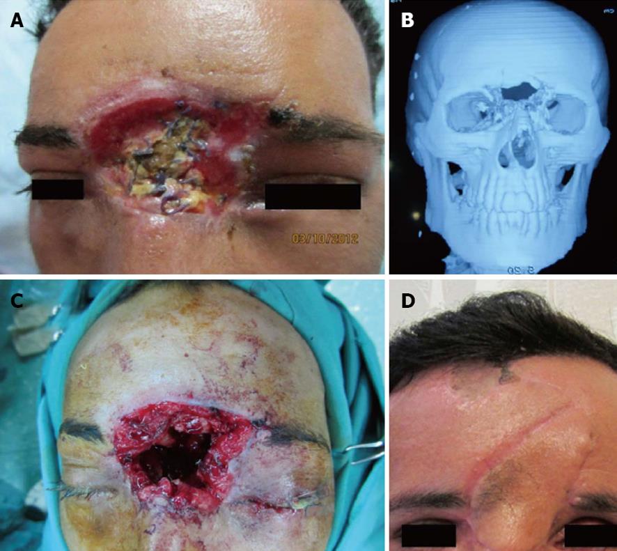
Figure 2 A 30-year old man with a gunshot wound to the upper face, with disruption of the forehead and frontal and ethmoid sinus and left eye to the base of the skull.
A: Before operation; B: The patient was treated with abdominal fat to obliterate the frontal sinus elsewhere before referral. Computed tomography scan before reconstruction; C: Intra operative view; D: Reconstruction with forehead flap and iliac bone graft (one month post operation).
Figure 3 A 23-year old man with a gunshot wound defect of the eyebrow.
A: Before operation; B: Early post operation; C: One year post operation.
- Citation: Ebrahimi A, Motamedi MHK, Nejadsarvari N, Kazemi HM. Management of missile injuries to the maxillofacial region: A case series. World J Stomatol 2013; 2(3): 62-66
- URL: https://www.wjgnet.com/2218-6263/full/v2/i3/62.htm
- DOI: https://dx.doi.org/10.5321/wjs.v2.i3.62


