Copyright
©The Author(s) 2017.
World J Respirol. Mar 28, 2017; 7(1): 29-34
Published online Mar 28, 2017. doi: 10.5320/wjr.v7.i1.29
Published online Mar 28, 2017. doi: 10.5320/wjr.v7.i1.29
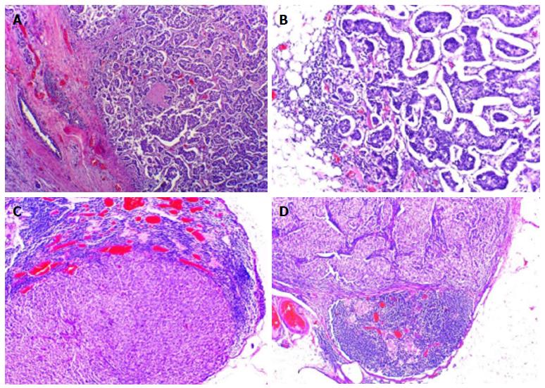
Figure 1 Invasive mammary carcinoma non-special type (A and B) (hematoxylin and eosin × 40-× 110), axillary lymph nodes metastases from mammary carcinoma (C and D) (hematoxylin and eosin × 40).
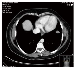
Figure 2 Spiculated nodular lesion with 18 mm localized in the left lower pulmonary lobe.
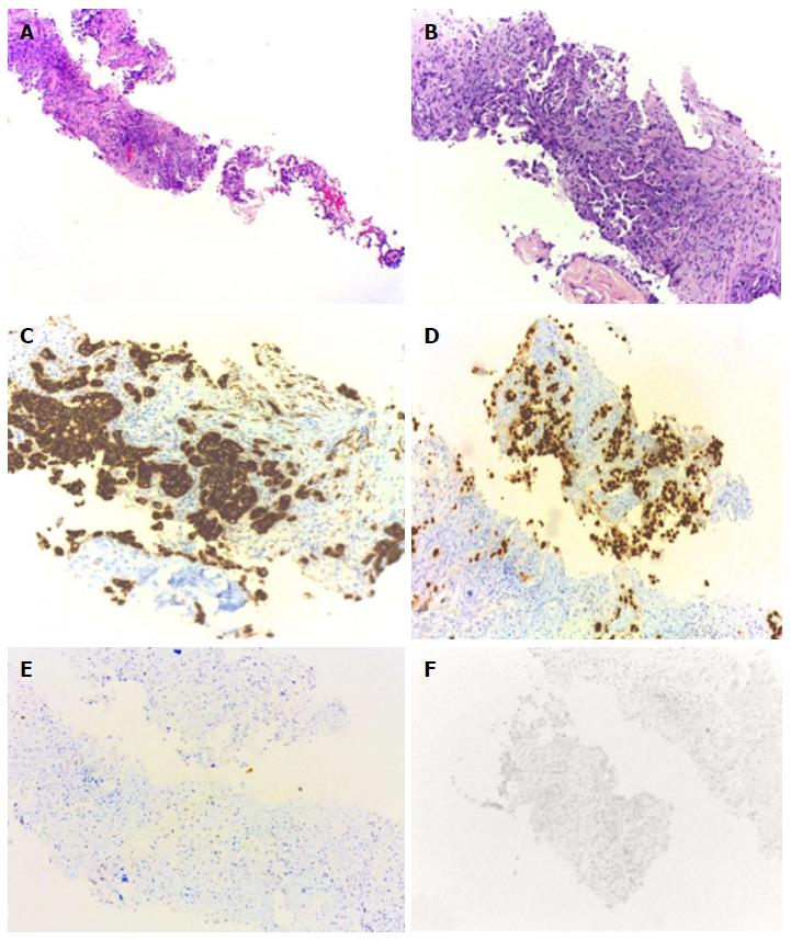
Figure 3 Transtoracic pulmonary biopsy.
Invasive adenocarcinoma (A and B) (HE × 40-× 100), Keratin 7 (C) and TTF1 (D) were positives. GATA 3 (E) and mammaglobin (F) were negatives. ER were negative (not shown). HE: Hematoxylin and eosin; ER: Estrogen receptor.
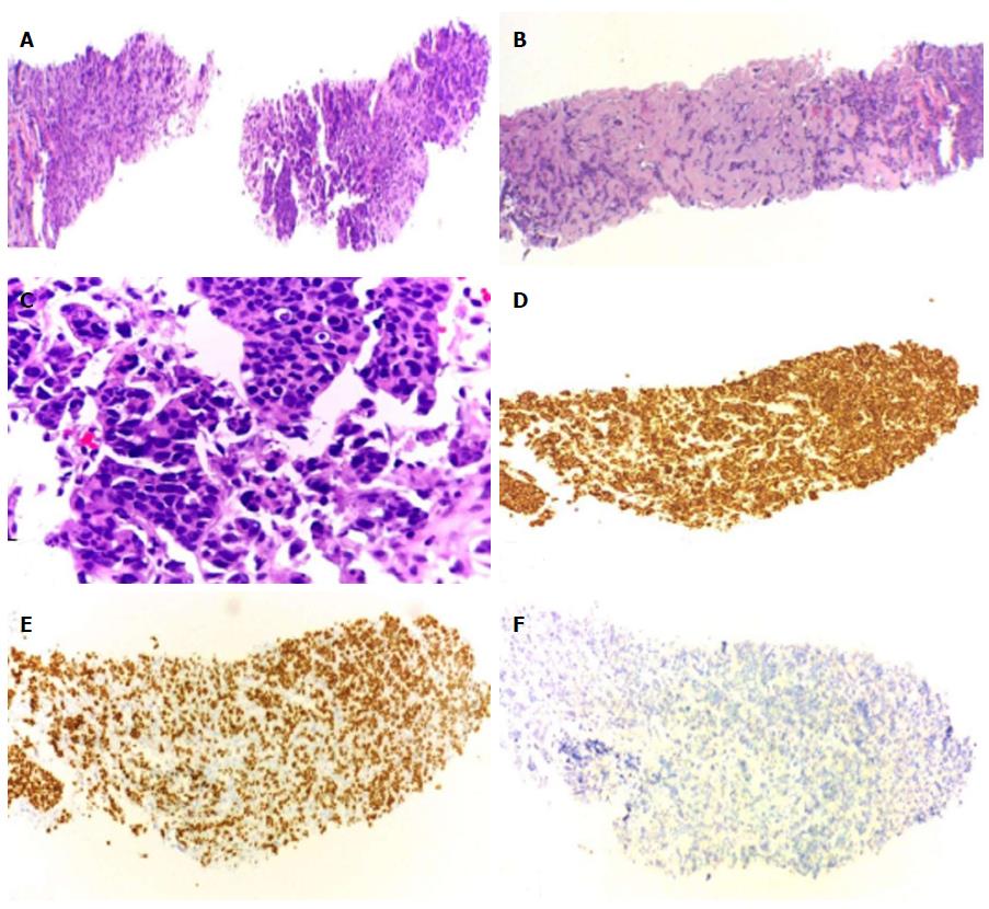
Figure 4 Left adrenal biopsy.
Solid-pattern carcinoma (A-C) (HE 40 ×-400 ×), Keratin 7 (D), GATA 3 (E) were positives. TTF1 was negative (F). ER positive and negative mammaglobin not shown. HE: Hematoxylin and eosin; ER: Estrogen receptor.
- Citation: de Macedo JE. Synchronous lung and breast cancer. World J Respirol 2017; 7(1): 29-34
- URL: https://www.wjgnet.com/2218-6255/full/v7/i1/29.htm
- DOI: https://dx.doi.org/10.5320/wjr.v7.i1.29


