Copyright
©The Author(s) 2015.
World J Respirol. Jul 28, 2015; 5(2): 126-134
Published online Jul 28, 2015. doi: 10.5320/wjr.v5.i2.126
Published online Jul 28, 2015. doi: 10.5320/wjr.v5.i2.126
Figure 1 Diagram summarizing the different functional domains of caveolin-1 protein.
Cav-1 contains seven known functional domains. It contains an oligomerization domain (OD), a caveolin scaffolding domain (CSD), a transmembrane domain (TMD), a caveolin inhibitory domain (CID) (eNOS, Src kinase and PKA), a terminal domain (TD), an N-terminal membrane association domain (N-MAD), and a C-terminal membrane association domain (C-MAD). P-133, 143, 156: Palmitoylation sites.
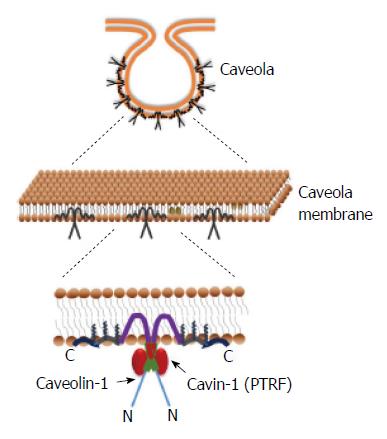
Figure 2 Structural organizations of a caveola, caveolin-1 and cavin-1.
Caveola are specialized lipid raft that are structurally maintained by caveolin-1 to form flask-shaped invaginations. In addition to these coat protein caveolin, caveolae contains an inner lining of adapter proteins called cavins, which regulate caveolin. PTRF: Polymerase I and transcript release factor.
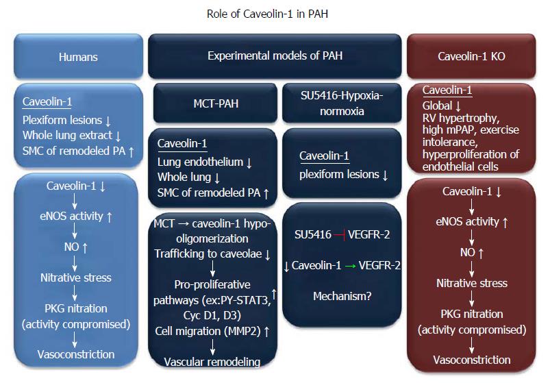
Figure 3 Schematic representation of alterations in caveolin-1 and the downstream pathways affected by caveolin-1 in human idiopathic pulmonary arterial hypertension and experimental models of pulmonary hypertension.
Cav-1 expression is decreased in the lung. Downstream signaling pathways that are affected by cav-1 are diverse in different animal models of pulmonary hypertension (PH) and in humans. However, they eventually lead to vasoconstriction, vascular remodeling and development of PH. PAH: Pulmonary arterial hypertension; VEGFR: Vascular endothelial growth factor receptor; Cav-1: Caveolin-1.
- Citation: Chettimada S, Yang J, Moon HG, Jin Y. Caveolae, caveolin-1 and cavin-1: Emerging roles in pulmonary hypertension. World J Respirol 2015; 5(2): 126-134
- URL: https://www.wjgnet.com/2218-6255/full/v5/i2/126.htm
- DOI: https://dx.doi.org/10.5320/wjr.v5.i2.126


