Copyright
©2012 Baishideng.
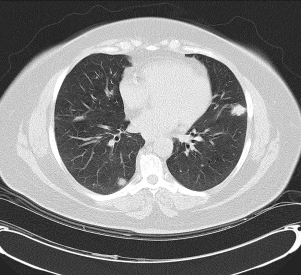
Figure 1 Computed tomography-scan of chest showing multiple bilateral pulmonary nodules of varying size.
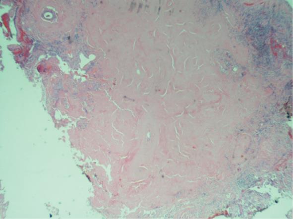
Figure 2 Nodular deposits of acellular, eosinophilic amyloid (Hematoxylin and eosin stain, × 40).
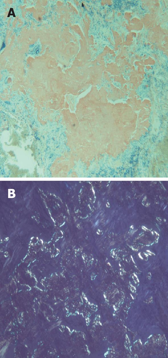
Figure 3 Amyloid deposits (Congo red stain, × 10) (A) and amyloid deposits under cross-polarized light demonstrating characteristic apple green birefringence (Congo red stain, × 40) (B).
- Citation: Bhavsar T, Huang Y, Gaughan C, Inniss S, Thomas R. Bilateral pulmonary nodular amyloidosis: A case report and review of the literature. World J Respirol 2012; 2(2): 6-8
- URL: https://www.wjgnet.com/2218-6255/full/v2/i2/6.htm
- DOI: https://dx.doi.org/10.5320/wjr.v2.i2.6


