Copyright
©2013 Baishideng.
World J Otorhinolaryngol. Feb 28, 2013; 3(1): 22-25
Published online Feb 28, 2013. doi: 10.5319/wjo.v3.i1.22
Published online Feb 28, 2013. doi: 10.5319/wjo.v3.i1.22
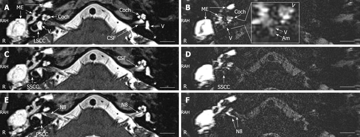
Figure 2 Magnetic resonance imaging of the inner ear acquired with a 3 Tesla machine.
In the T2-weighted images (A, C, E), intense signals in the perilymph and endolymph of the inner ear and cerebrospinal fluid (CSF) surrounding the eighth nerve (N8) and the middle ear cavity (ME) were demonstrated. In the heavily T2-weighted 3-dimensional fluid-attenuated inversion recovery magnetic resonance images, evident enhancement by gadolinium-tetraazacyclododecane-tetraacetic acid (Gd-DOTA) was detected in the middle ear cavity and the perilymphatic compartments of the cochlea. Suspected endolymphatic hydrops was indicated by the enlarged scala media at the basal turn [arrowhead in the enlarged window of (B)]. In general, the Gd-DOTA uptake in the vestibule was weak, and endolymphatic hydrops became obvious in the vestibulum (V) and ampulla of the semicircular canal (Am) [enlarged window of (B)]. Gd-DOTA uptake in the perilymph of superficial semicircular canal was detected in the diseased ear (D). No uptake of Gd-DOTA was demonstrated neither in the lateral semicircular canal nor in the posterior semicircular canal. The N8 (in F) on the diseased side showed significant enhancement. LSCC: Lateral semicircular canal; PSCC: Posterior semicircular canal; SSCC: Superior semicircular canal.
- Citation: Zou J, Pyykkö I. Endolymphatic hydrops in Meniere’s disease secondary to otitis media and visualized by gadolinium-enhanced magnetic resonance imaging. World J Otorhinolaryngol 2013; 3(1): 22-25
- URL: https://www.wjgnet.com/2218-6247/full/v3/i1/22.htm
- DOI: https://dx.doi.org/10.5319/wjo.v3.i1.22


