Copyright
©The Author(s) 2019.
World J Otorhinolaryngol. Apr 27, 2019; 8(1): 4-11
Published online Apr 27, 2019. doi: 10.5319/wjo.v8.i1.4
Published online Apr 27, 2019. doi: 10.5319/wjo.v8.i1.4
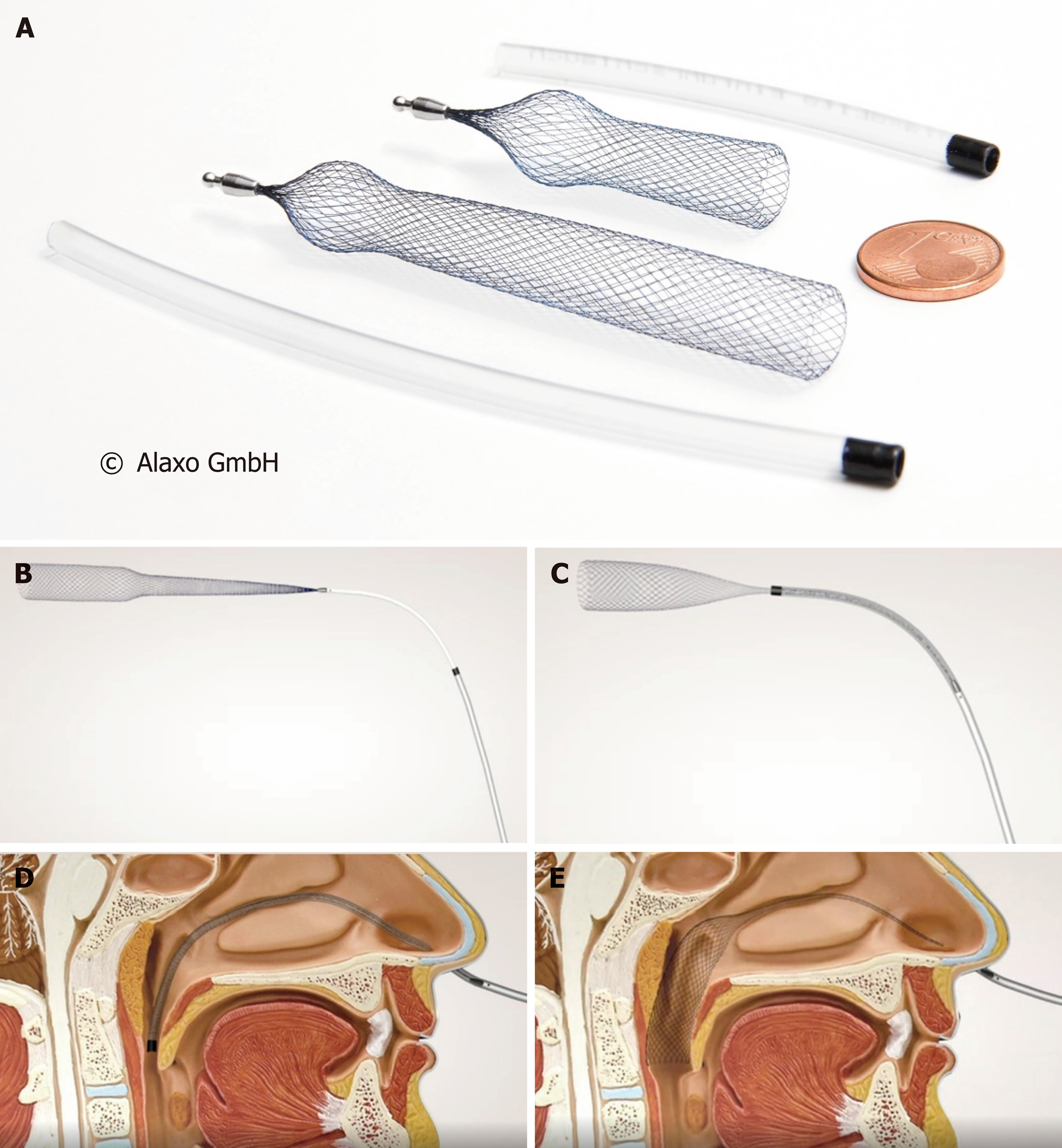
Figure 1 AlaxoLito (A) and AlaxoLito Plus (C-E) Nasal Stents with insertion tube.
Insertion method: B and C: The stent is placed inside the insertion tube, which allows for easy insertion; D: Once the insertion tube is placed into the nasopharynx; E: It is withdrawn, allowing the stent to open inside the nasal cavity.
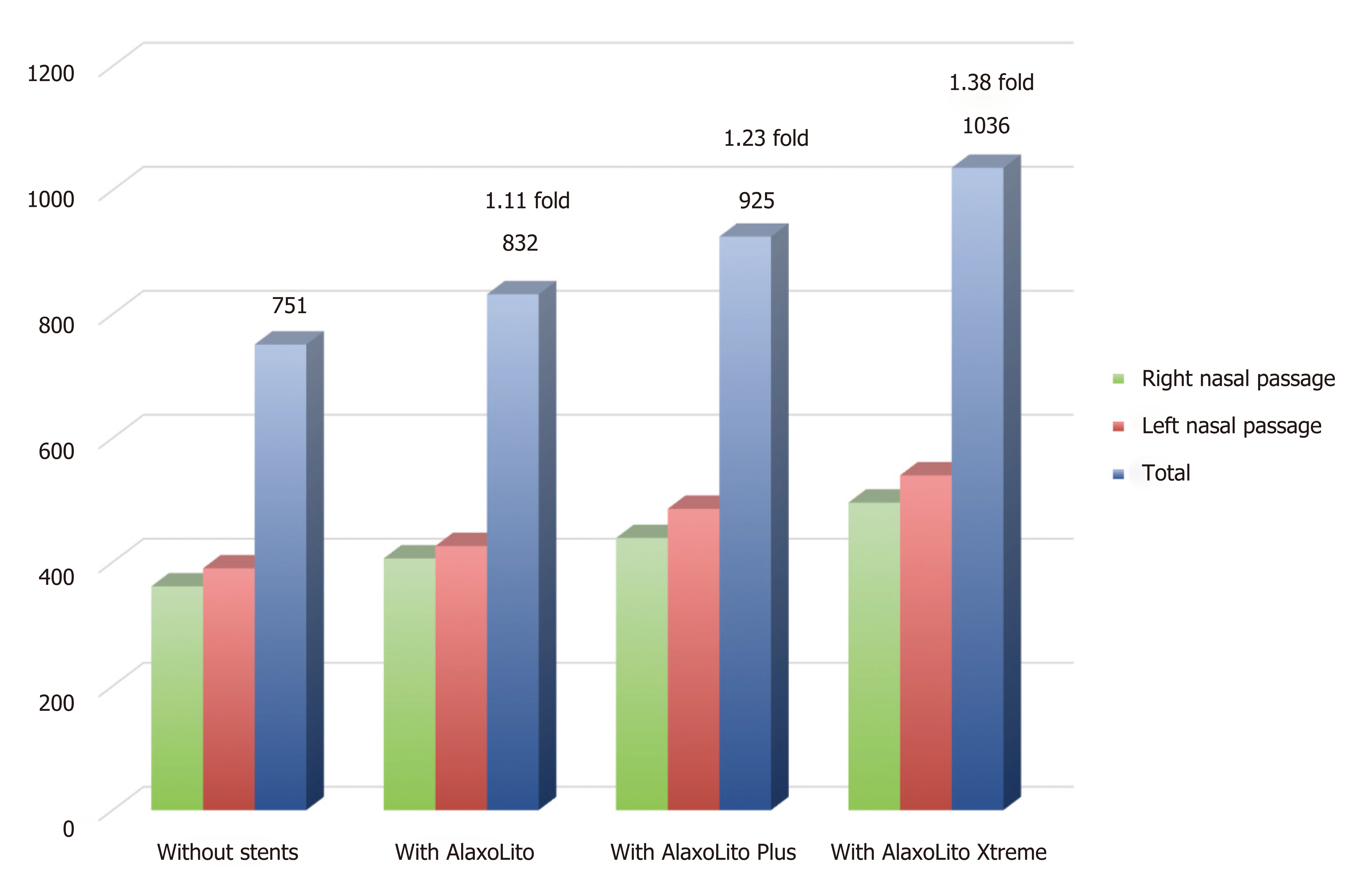
Figure 2 Rhinomanometric data without and with stents.
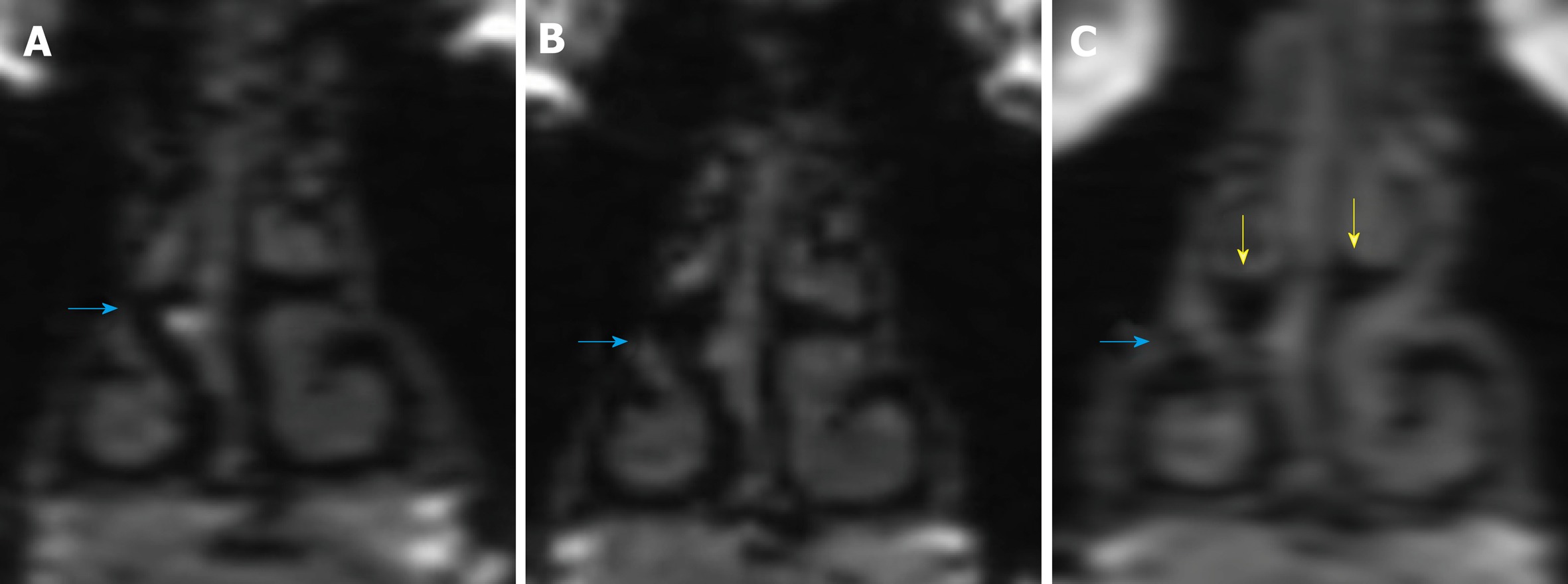
Figure 3 Comparison of coronal magnetic resonance imaging of nasal passages prior to and 1 year post regular stent use.
A: Magnetic resonance imaging (MRI) from December 2015 prior to regular use of stents, without stents; B and C: MRI from June 2017 one year post regular use of AlaxoLito Plus stent (B: without stents in situ; C: with AlaxoLito Plus in situ).
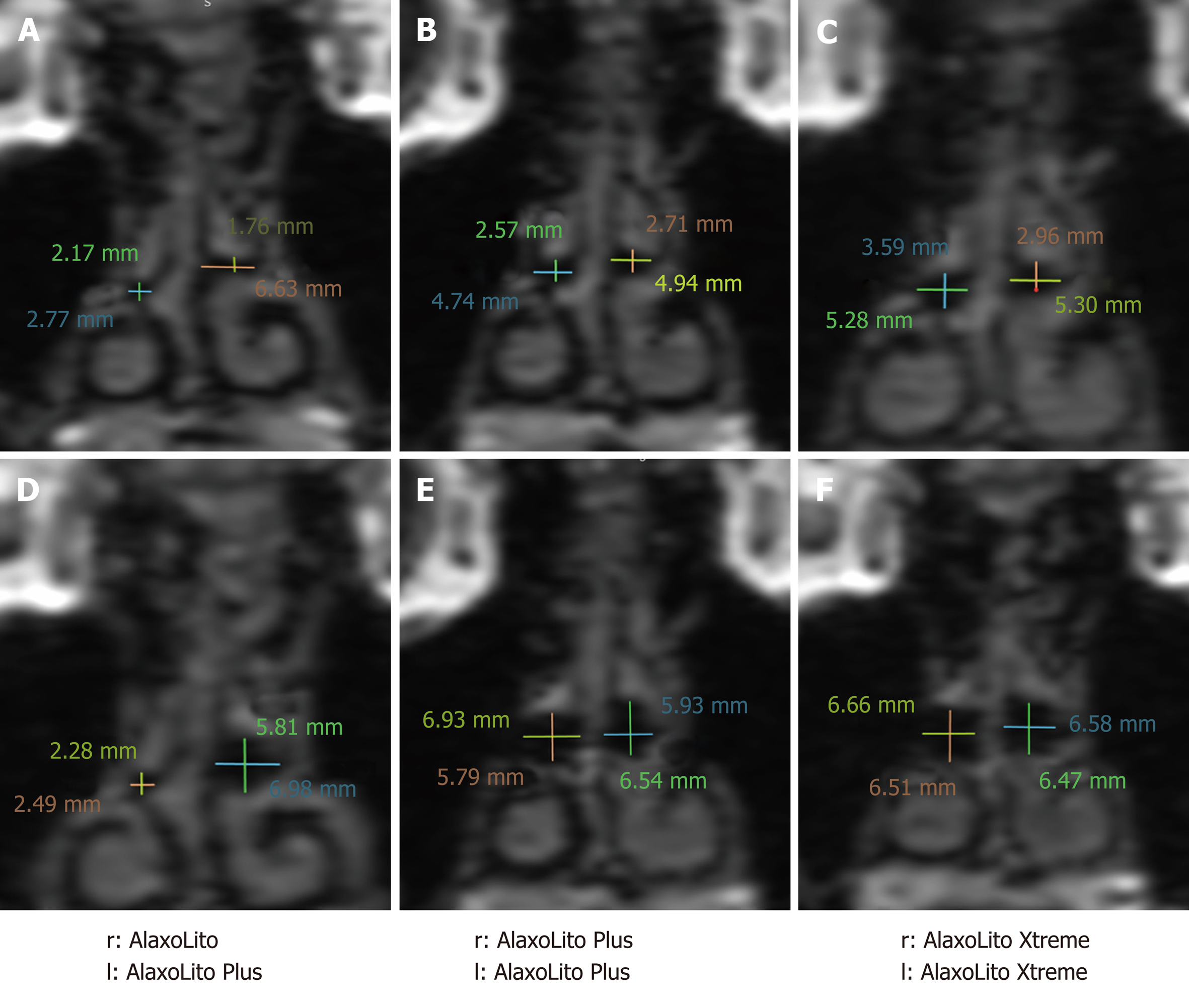
Figure 4 Coronal magnetic resonance imaging showing measurements of middle nasal passage without stents (A-C) and with stents in situ (D-F).
A and D: Magnetic resonance imaging (MRI) from December 2015 prior to regular use of stents; B and E: MRI from June 2017 one year post regular use of AlaxoLito Plus; C and F: MRI from September 2018, 3.5 mo post regular use of AlaxoLito Xtreme.
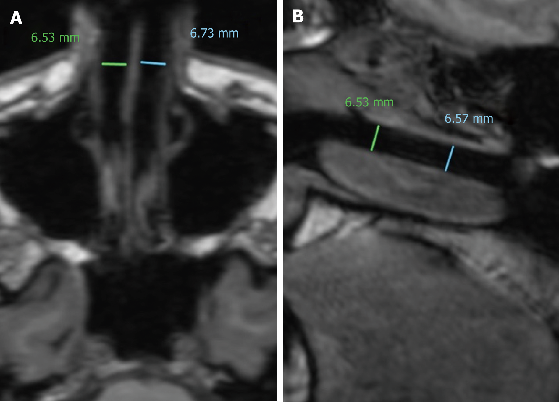
Figure 5 Axial (A) and sagittal (B) magnetic resonance imaging (from September 2018) showing measurements of middle nasal passage with AlaxoLito Xtreme stents in situ.
- Citation: Zhang H, Kotecha B. Effect of intranasal stents (AlaxoLito, AlaxoLito Plus and AlaxoLito Xtreme) on the nasal airway: A case report. World J Otorhinolaryngol 2019; 8(1): 4-11
- URL: https://www.wjgnet.com/2218-6247/full/v8/i1/4.htm
- DOI: https://dx.doi.org/10.5319/wjo.v8.i1.4


