Copyright
©The Author(s) 2016.
World J Otorhinolaryngol. Feb 28, 2016; 6(1): 13-18
Published online Feb 28, 2016. doi: 10.5319/wjo.v6.i1.13
Published online Feb 28, 2016. doi: 10.5319/wjo.v6.i1.13
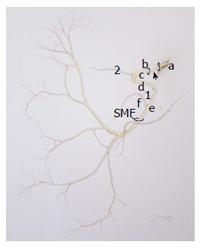
Figure 1 Line drawing of the facial nerve.
The nerve takes a very tortuous course through the temporal bone within the Fallopian canal before exiting the skull base at the SMF. Any portion, intra or extratemporal, can be affected by a facial nerve schwannoma. a: Nerve as it exits the brainstem and crosses the cerebellopontine angle; b: Labyrinthine segment; c: Geniculate ganglion; d: Tympanic segment; e: Mastoid segment; f: Chorda tympani nerve; arrow: Intracanilicular segment. 1Nerve to stapedius; 2Greater superficial petrosal nerve. (Joshua M Klein, MSM I artist). SMF: Stylomastoid foramen.
Figure 2 Radiographic images of facial nerve schwannoma.
A: Contrasted coronal T1 weighted MRI of right intracanilicular FNS. The patient was presumed to have a vestibular schwannoma but the tumor arose from the FN. The patient underwent tumor debulking with HB grade 1 at follow-up; B: Contrasted T1 weighted coronal MRI of a large FNS arising from the geniculate ganglion with significant extension into the middle fossa and the middle ear. The patient underwent a translabryinthine resection with cable grafting; C: Post contrast axial T1 weighted MRI demonstrating a FNS involving multiple levels of the facial nerve. This is a typical appearance of the “beads on a string” pattern of tumor growth (arrrowheads); D: Axial bone window high resolution computer tomography of the temporal bone demonstrating a large FNS of the mastoid segment causing erosion of both the bony ear canal (arrowhead) and the posterior fossa plate. MRI: Magnetic resonance imaging; FNS: Facial nerve schwannoma; HB: House-Brackmann.
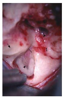
Figure 3 Intraoperative photograph of a left subtemporal/middle cranial fossa approach to the facial nerve.
The middle fossa approach to the internal auditory canal is appropriate for decompression of the bony canal surrounding the nerve. Access to the upper tympanic segment (proximal to the cochleariform process) is achievable via this approach. 1Geniculate ganglion. Solid line represents the entrance of the bony facial canal; Solid black arrow: Blue-line of superior semicircular canal; Black arrowhead: Fundus of the internal auditory canal decompressed; White arrow: Labyrinthine segment; White arrowhead: Tympanic segment.
- Citation: Makadia L, Mowry SE. Management of intratemporal facial nerve schwannomas: The evolution of treatment paradigms from 2000-2015. World J Otorhinolaryngol 2016; 6(1): 13-18
- URL: https://www.wjgnet.com/2218-6247/full/v6/i1/13.htm
- DOI: https://dx.doi.org/10.5319/wjo.v6.i1.13


