Copyright
©The Author(s) 2015.
World J Otorhinolaryngol. Feb 28, 2015; 5(1): 30-36
Published online Feb 28, 2015. doi: 10.5319/wjo.v5.i1.30
Published online Feb 28, 2015. doi: 10.5319/wjo.v5.i1.30
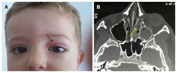
Figure 1 Swelling and redness at the level of the left upper eyelid (A); the computed tomography-scan shows a fulfilling of the ethmoïd cells and the left maxillary sinus (B).
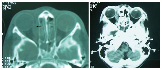
Figure 2 Bilateral ethmoiditis (A and B) complicated by a subperiosteal abscess (arrows).
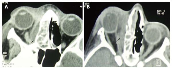
Figure 3 Right ethmoiditis complicated by intraorbital abscess (A and B).
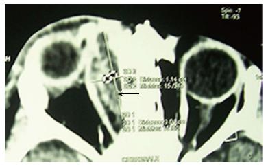
Figure 4 Intraorbital abscess displacing the ocular globe anteriorly and laterally (arrow).
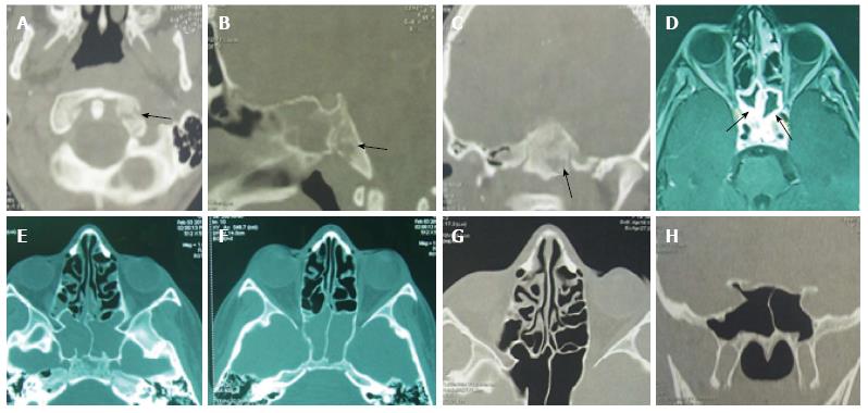
Figure 5 Sphenoiditis with bony lysis and a notable regression of the inflammatory lesions in a 9-year-old child.
Sphenoiditis in a 9-year-old child with bony lysis in computed tomography-scan (A, B and C) and magnetic resonance imaging (D). The regression of the inflammatory lesions after sphenoidotomy and antibiotherapy was partial after 3 wk (E and F) and complete after 5 wk (G and H).
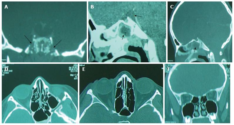
Figure 6 Sphenoiditis with meningitis in an 11-year-old child treated by sphenoidotomy and intravenous antibiotherapy.
Note the lytic lesions of the sphenoid bone (A-C). The computed tomography-scan, performed after 1 mo shows a complete regression of the inflammatory signs (D-F).
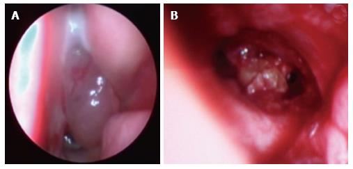
Figure 7 Sphenoiditis have benefited from endoscopic sphenoidotomy with intravenous antibiotherapy.
Nasal endoscopy showing an inflammatory polyp with purulent secretions coming from the right sphenoid sinus (A). The sphenoidotomy revealed an inflammatory granuloma into the sphenoid cavity (B).
- Citation: Mardassi A, Mathlouthi N, Mbarek H, Halouani C, Mezri S, Zgolli C, Chebbi G, Mhamed RB, Akkari K, Benzarti S. Complicated sinusitis in children: 18 cases report. World J Otorhinolaryngol 2015; 5(1): 30-36
- URL: https://www.wjgnet.com/2218-6247/full/v5/i1/30.htm
- DOI: https://dx.doi.org/10.5319/wjo.v5.i1.30


