Copyright
©The Author(s) 2015.
World J Otorhinolaryngol. Feb 28, 2015; 5(1): 21-29
Published online Feb 28, 2015. doi: 10.5319/wjo.v5.i1.21
Published online Feb 28, 2015. doi: 10.5319/wjo.v5.i1.21
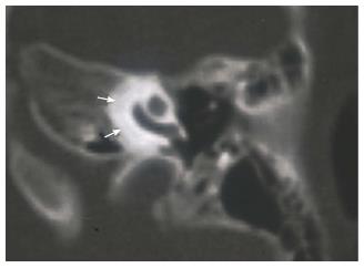
Figure 1 CT appearance of cochlear otosclerosis.
Note for perilabyrinthine decalcification (marked with arrows).
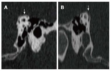
Figure 2 Bilateral superior semicircular canal dehiscence of A and B (marked with arrows).
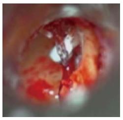
Figure 3 Revision stapes surgery due to incus necrosis.
Bone cement was used to reconstruct the incus. Titanium prosthesis was placed over the fixed incus.
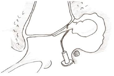
Figure 4 Ossicular reconstructions between the mobile malleus and stapes footplate fenestra if incus is not available due dislocation or extensive necrosis.
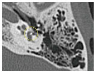
Figure 5 Temporal bone tomography with long prosthesis inside the vestibule (marked with yellow circle).
- Citation: Yetiser S. Revision surgery for otosclerosis: An overview. World J Otorhinolaryngol 2015; 5(1): 21-29
- URL: https://www.wjgnet.com/2218-6247/full/v5/i1/21.htm
- DOI: https://dx.doi.org/10.5319/wjo.v5.i1.21


