Copyright
©2014 Baishideng Publishing Group Inc.
World J Otorhinolaryngol. Aug 28, 2014; 4(3): 12-16
Published online Aug 28, 2014. doi: 10.5319/wjo.v4.i3.12
Published online Aug 28, 2014. doi: 10.5319/wjo.v4.i3.12
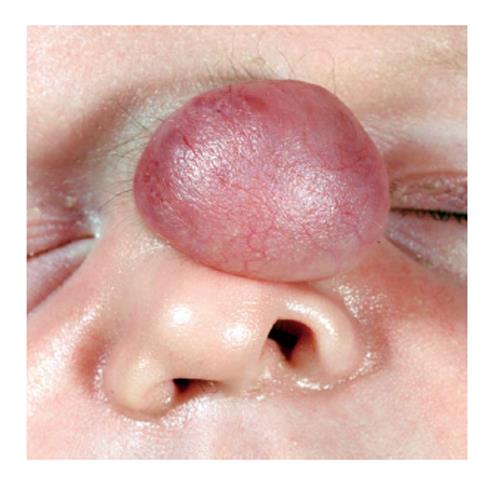
Figure 1 Clinical image of Nasal glioma in a neonate (original image with permission)[13].
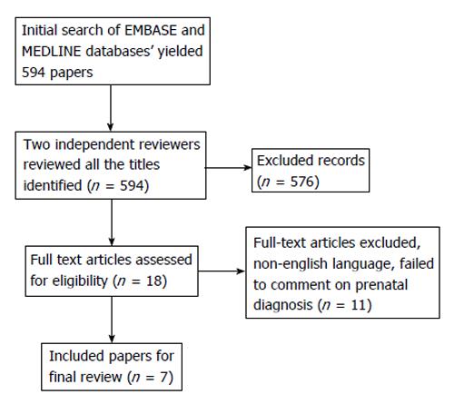
Figure 2 PRISMA flow diagram for inclusion/exclusion.
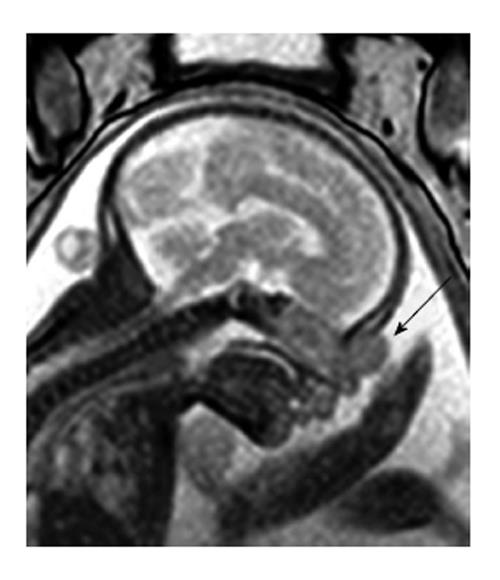
Figure 3 Sagittal foetal magnetic resonance imaging in utero, identifying nasal lesion (arrow).
Original image with permission[13].
- Citation: Fox R, Okhovat S, Beegun I. Prenatal diagnosis and management of nasal glioma. World J Otorhinolaryngol 2014; 4(3): 12-16
- URL: https://www.wjgnet.com/2218-6247/full/v4/i3/12.htm
- DOI: https://dx.doi.org/10.5319/wjo.v4.i3.12


