Copyright
©2012 Baishideng Publishing Group Co.
World J Otorhinolaryngol. Feb 28, 2012; 2(1): 1-7
Published online Feb 28, 2012. doi: 10.5319/wjo.v2.i1.1
Published online Feb 28, 2012. doi: 10.5319/wjo.v2.i1.1
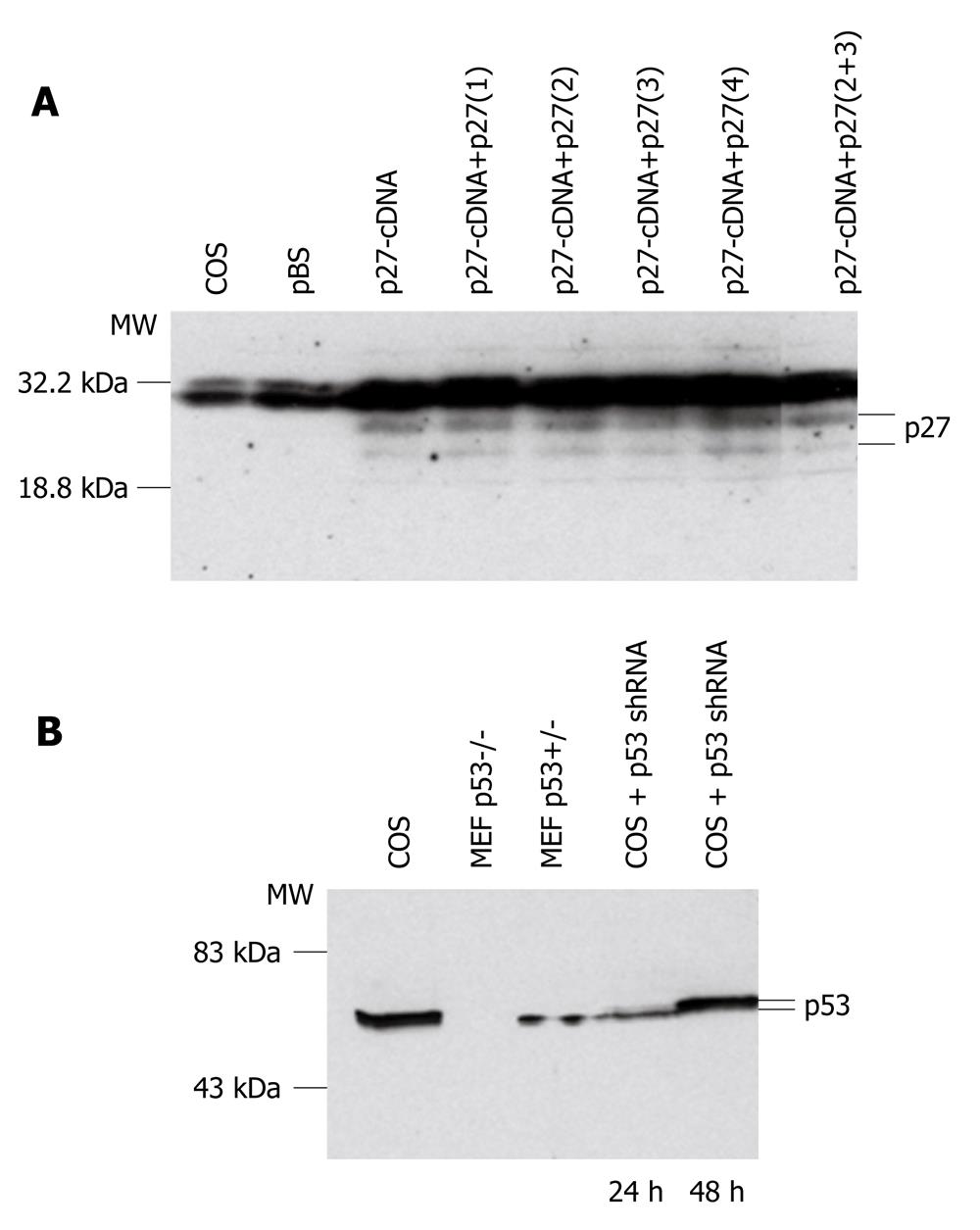
Figure 1 Short hairpin RNA testing in COS cells.
A: Western blot analysis with anti-p27Kip1 antibody. Proteins were extracted from untransfected COS cells and COS cells transfected with Bluescript (pBS), p27Kip1 cDNA (p27-cDNA) or p27Kip1 cDNA together with 4 different p27Kip1 short hairpin RNA (shRNA) constructs, p27(1-4). p27Kip1 specific bands are marked on the right side. A decrease in p27Kip1 level was observed with p27(2), p27(3) and p27(2+3) after 24 h of transfection; B: Western blot analysis with anti-p53 antibody. Proteins were extracted from untransfected COS cells (COS), p53 null cells (p53-/-), p53 heterozygous null cells (p53+/-), COS cells transfected with p53 shRNA. Cells were collected 24 h or 48 h after transfection. p53 specific bands are marked on the right side. A decrease in p53 level was detected after 24 h of transfection. Kaleidoscope molecular weight (MW) standards are marked on the left side. MEF: Mouse embryonic fibroblasts.
Figure 2 Schematic representation of used adeno-associated virus constructs.
The constructs are not drawn in scale. ITR: Adeno-associated virus inverted terminal repeats; CMV: Human cytomegalovirus promoter; U6: U6-promoter; EGFP: Enhanced green fluorescent protein coding sequence; shRNA: Small hairpin RNA; 1: shp27Kip1(2)/shp27Kip1(3); 2: shp53; 3: shp53/shp27Kip1(2)/shp27Kip1(3); WPRE: Woodchuck hepatitis virus post-transcriptional regulatory element; pA: SV40 polyadenylation signal.
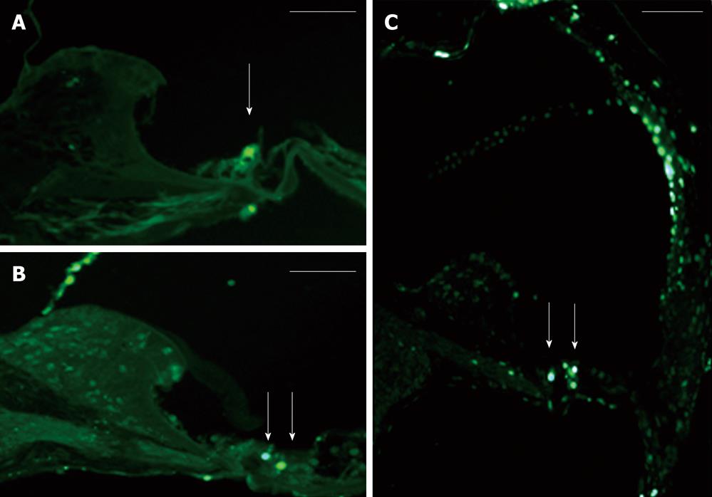
Figure 3 Adeno-associated virus-short hairpin RNAs in mouse cochlea.
A: EGFP expression in mouse cochlea organ of Corti cotransduced with adeno-associated virus (AAV)-EGFP and AAV-short hairpin RNA (shRNA) (p27Kip1+p53). Arrow shows EGFP-positive cell; FITC filter, Scale bar 200 μm; B: Uninjected control ear form the AAV-shRNA (p27Kip1+p53) injected animal; Arrows point to apoptotic cells in organ of Corti, Scale bar 200 μm; C: NaCl injected animal’s ear; Arrows point to apoptotic cells in organ of Corti. Scale bar 200 μm.
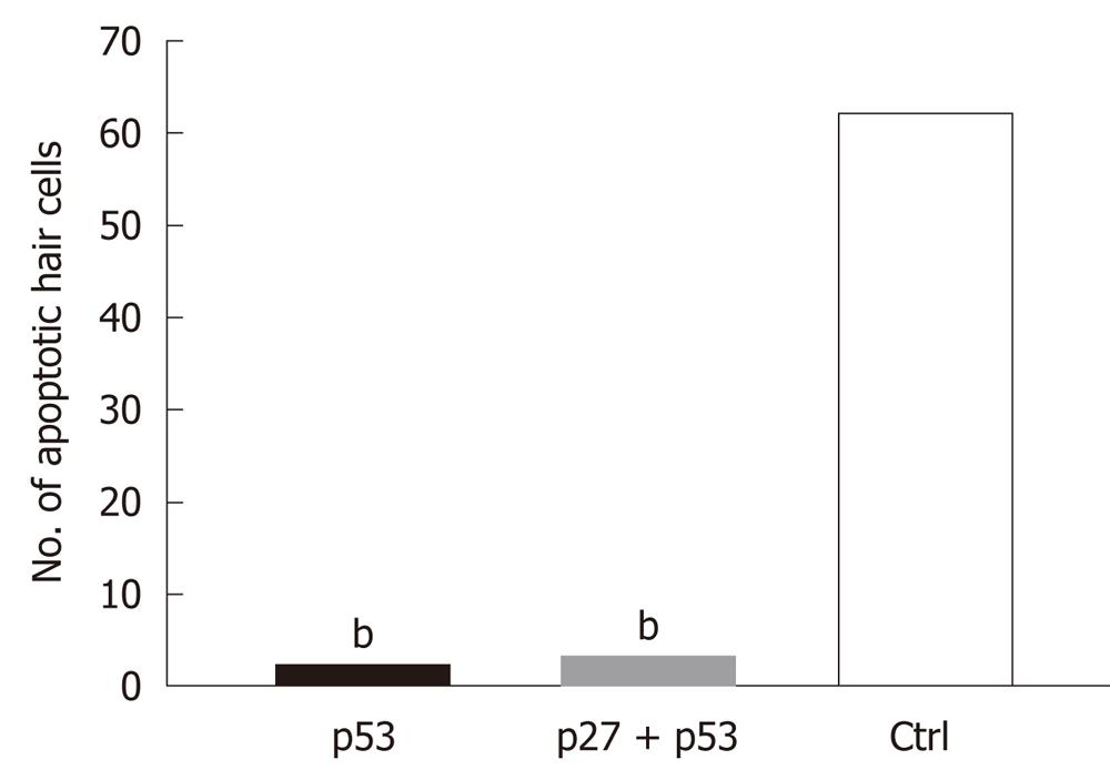
Figure 4 Number of apoptotic cells in the mouse cochlea after kanamycin treatment and injections with adeno-associated virus-p53-short hairpin RNA and adeno-associated virus-p27 + p53-short hairpin RNA constructs.
Differences in the level of apoptosis between adeno-associated virus (AAV)-short hairpin RNA (shRNA) and saline injected cochleae were statistically significant, bP < 0.001. AAV-p53-shRNA vs saline P = 0.00014; AAV-p27 + p53-shRNA vs saline P = 0.0011.
- Citation: Pietola L, Jero J, Jalkanen R, Kinnari TJ, Jero O, Frilander M, Pajusola K, Salminen M, Aarnisalo AA. Effects of p27Kip1- and p53- shRNAs on kanamycin damaged mouse cochlea. World J Otorhinolaryngol 2012; 2(1): 1-7
- URL: https://www.wjgnet.com/2218-6247/full/v2/i1/1.htm
- DOI: https://dx.doi.org/10.5319/wjo.v2.i1.1


