Copyright
©The Author(s) 2023.
World J Otorhinolaryngol. May 9, 2023; 10(2): 30-35
Published online May 9, 2023. doi: 10.5319/wjo.v10.i2.30
Published online May 9, 2023. doi: 10.5319/wjo.v10.i2.30
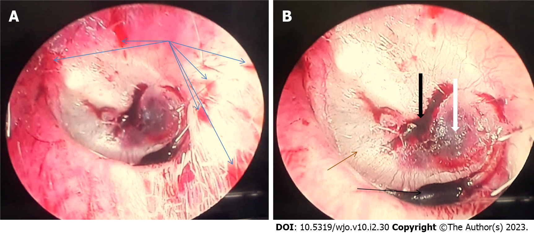
Figure 1 Shows multiple bleeding points (blue arrows) over the left external ear canal, two blood clots over the eardrum (black arrows), hemotympanum in the posterior part of the tympanic cavity (white arrow), absent cone of light, and dull-looking tympanic membrane (brown arrow).
A: A little bit far away from the tympanic membrane; B: Closer to the eardrum.
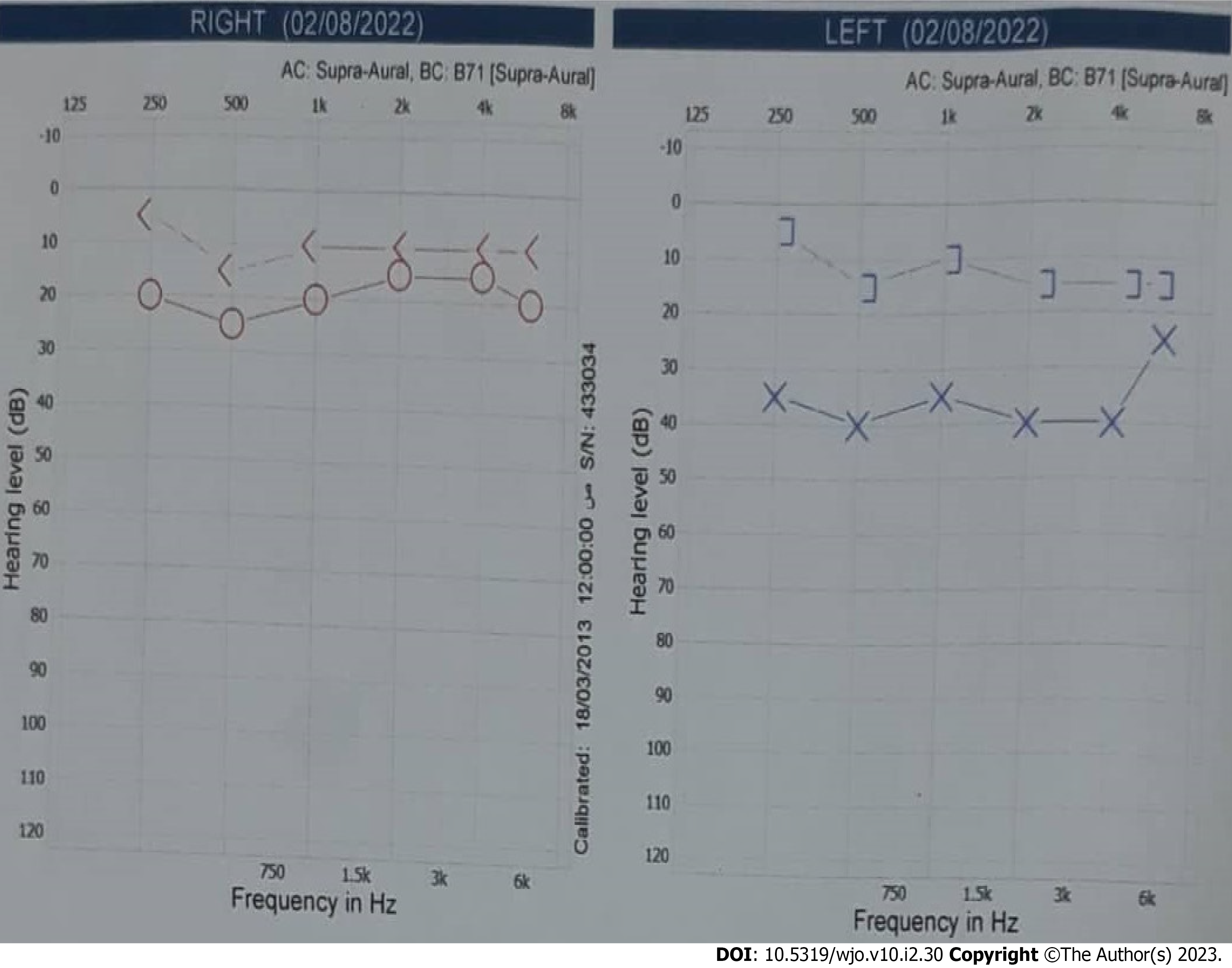
Figure 2 Pure tone audiogram of the 27-year-old man shows conductive hearing loss of 37 dB in the left ear.
Hz: Hertz; dB: Decibel; Circle: Air conduction hearing threshold at a specific frequency of the right ear; X: Air conduction hearing threshold at a specific frequency of the left ear; <: Bone conduction hearing threshold at a specific frequency of the right ear with masking of the left ear; ]: Bone conduction hearing threshold at a specific frequency of the left ear.
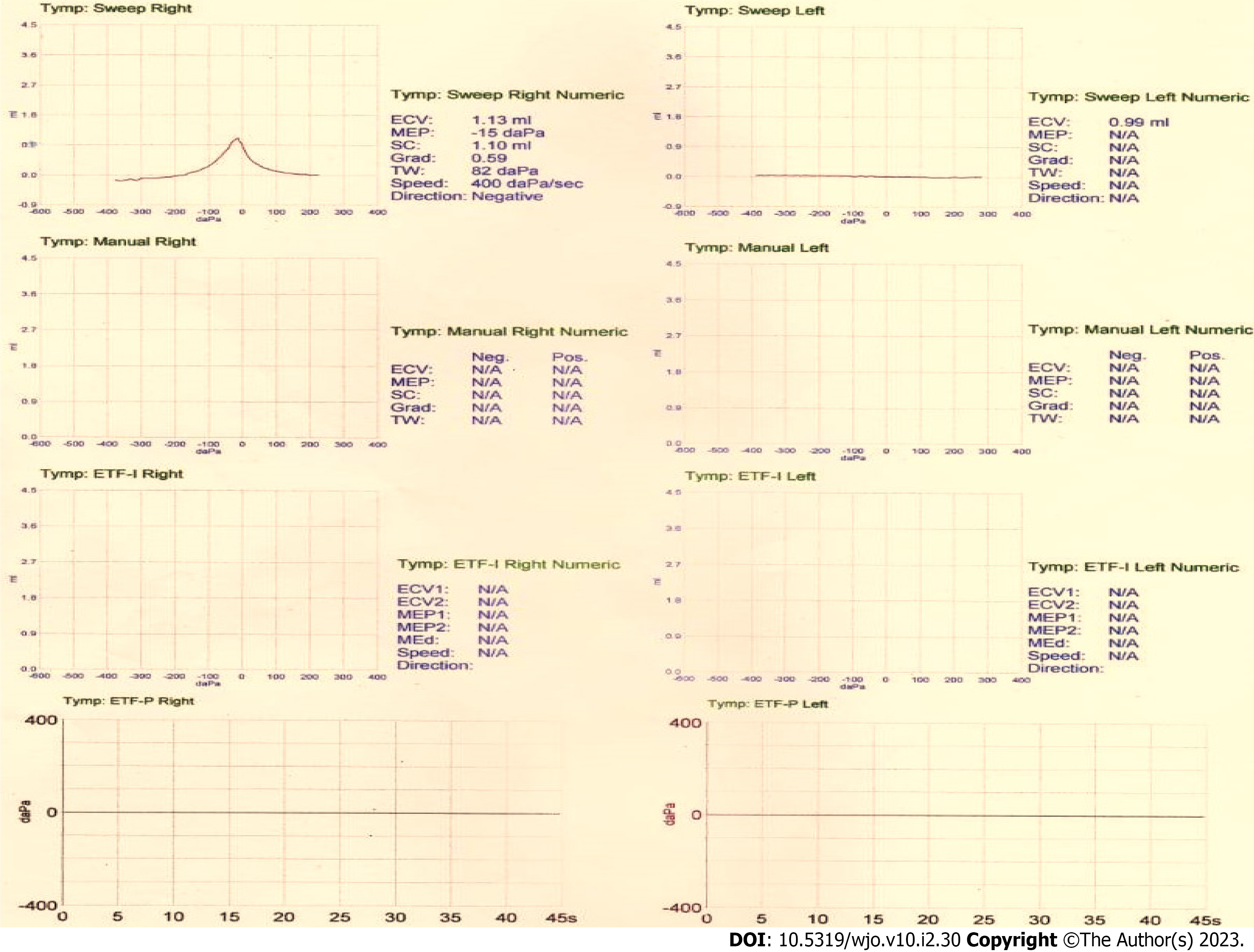
Figure 3 Tympanogram of the 27-year-old man shows type A on the right ear and type B (flat curve) on the left ear.
The left external canal volume is 0.99 mL.
- Citation: Al-Ani RM. Wet cupping (Al-hijamah) as a strange cause of ear trauma: A case report. World J Otorhinolaryngol 2023; 10(2): 30-35
- URL: https://www.wjgnet.com/2218-6247/full/v10/i2/30.htm
- DOI: https://dx.doi.org/10.5319/wjo.v10.i2.30


